Trichorrhexis invaginata
Microscopic examination of hair shaft: bamboo hair. Hair shaft with multiple knots resembling the ball-and-cup joint of bamboo
Dermoscopy: nodular swellings at irregular intervals, ball-shaped knots similar to match sticks, and hair breakages
Prenatal chorionic villus testing for SPINK5
Monilethrix
Microscopic examination of hair shaft: beaded hair. Hair shaft with elliptical nodes of normal hair and are medullated, regularly separated by internodes which are narrow, devoid of medulla, and are the site of fracture
Dermoscopy: beading and breakage at the internode level, with affected hairs bending in different directions
Menkes kinky hair syndrome
Microscopic examination of hair shaft: pili torti. The shafts are flattened and present twisting through 180° at irregular intervals
Dermoscopy: pili torti
Labs for serum copper, ceruloplasmin, and urine homovanillic/vanillylmadelic acid ratio
Prenatal diagnosis: placental copper levels are threefold to fivefold higher in pregnancies
Trichothiodistrophy
Microscopic examination of hair shaft: tiger tail hair. Under polarized microscopy the affected hair has a banded or “tiger tail” pattern because of alternating birefringence due to the presence of transverse dark and bright bands. Analyze proximal shaft to avoid changes due to weathering
Dermoscopy: useful to select the hairs to be examined under the microscope, as they present a dishomogeneous structures resembling grains of sand
For Treatment
There is no specific treatment for hair shaft fragility except for gentle handling of hair to minimize breakage. In most patients fragility improves with aging. Early identification and treatment of patients with Menkes kinky hair disease is paramount. By administering copper injections early, longer-term survival approaches 92 % [1]. Parenteral copper can prevent development of neurological abnormalities in some patients, but response to treatment depends on specific ATP7A mutations.
Trichorrhexis Invaginata (Bamboo Hair)
Clinical Features
The hair shaft shows multiple knots along its length. The knots consists of a proximal cup-shaped portion and a distal ball-shaped portion resembling the ball and cup joint of bamboo. Hair breakage occurs at the nodes. Hair of patients with trichorrhexis invaginata is dry, dull, fragile, and short due to hair breakage. Trichorrhexis invaginata frequently affects eyelashes, eyebrows and secondary sexual hair. The eyebrows usually show multiple bamboo nodes, and may present the abnormality even when the scalp hair, which improves with age, appears normal [2].
Trichorrhexis invaginata may be isolated, but most commonly is part of Netherton disease (MIM 256500). A rare autosomal recessive genodermatosis, which combines ichthyosis, bamboo hair and atopic dermatitis [3].
Monilethrix (Beaded Hair; MIM158000, 177750, 252700)
Clinical Features
The hair is dull, fragile, breaks easily at 0.5–2.5 cm from the scalp, and breakage is prominent in the nape and occipital areas (Fig. 7.1a). The severity of the alopecia may vary considerably from almost complete hair loss to very mild thinning. Even members of the same family can be differently affected. Follicular keratosis of the affected scalp and keratosis pilaris are typical.
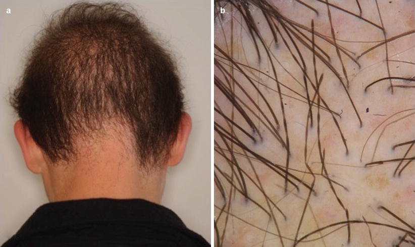
Fig. 7.1
(a, b) Monilethrix. (a) Diffuse hair thinning with pronounced loss in the occipital region. Hair is dull and short. (b) Characteristic beading of hair seen on dermoscopy
Monilethrix does not affect lanugo hair and usually becomes evident with growing of mature hair. At birth the scalp hair is either normal or absent; in the latter case the scalp has a shaved appearance. Scraping of the scalp may show the typical beads resulting from broken monilethrix hairs. Hair fragility improves with age. Monilethrix can occasionally affect eyebrows and eyelashes.
Monilethrix is most commonly inherited as an autosomal dominant condition with variable expression. Several mutations in the human basic hair keratins (hHb1 and hHb6) have been reported [4, 5] with the most frequent mutation E413K occurring in hHb6 [6]. Linkage to the type II keratin cluster is on 12q13. Recessive monilethrix is rare and has been linked to mutations in desmoglein 4 (DSG4) [7].
Investigations Recommended
For Diagnosis
Diagnosis is made by clinical examination utilizing dermoscopy and in some instances microscopic examination of the hair shaft (see Table 7.1).
Microscopic examination of the hair shaft: the hair shaft has a beaded appearance due to the presence of elliptical nodes, which have the diameter of normal hair and are medullated, regularly separated by internodes which are narrow, devoid of medulla and are the site of fracture.
Menkes Kinky Hair Syndrome (MIM 309400)
Clinical Features
The hair is fine, silver or white, fragile and has a kinky/wiry appearance. The abnormal hair is usually not present at birth, but becomes evident around 4–5 months of age. Menkes kinky hair syndrome is an X-linked neurodegenerative disorder of copper metabolism, which combines pili torti and progressive neurological dysfunction [9]. The Menkes gene mutation has been mapped to Xq13.3 affecting the ATP7A gene. Pili torti in the hair of the mother of a patient with Menkes kinky hair disease is considered definitive proof of her status as a gene carrier.
Trichothiodystrophy (MIM 601675)
Clinical Features
Trichothiodystrophy is due to a reduction in the sulfur and cystine content of hair. The hair is brittle, sparse, dry, unruly and short. Eyebrows and eyelashes can also be affected. Brittle nails are often associated with trichothiodystrophy. The condition is autosomal recessive. The presence of associated features allows for distinguishing several syndromes (see Table 7.2). Photosensitivity, with mutations of the XPD, XPB or TTDA gene is present in about 50 % of cases. However, there is no predisposition to skin cancers in these patients [10].
Table 7.2
Classification of trichothiodistrophy
Type | MIM | Findings | Eponym/acronym |
|---|---|---|---|
A | Hair ± nail involvement | ||
B | 211390 | Hair ± nails + mental retardation | Sabinas |
C | 275550 | Hair, mental retardation, folliculitis, retarded bone age ± caries and ± nail involvement | Pollitt |
D | 234050 | Brittle hair, infertility, developmental delay, short stature and ± nail involvement | BIDS |
E | 242170 | Ichthyosis + BIDS. Brittle hair, ± nail involvement, mental retardation, short stature, ± decreased gonadal function, ± lenticular opacities/cataracts, failure to thrive, microcephaly ± ataxia, ± calcifications of the basal ganglia, erythroderma and scale | Tay + BIDS |
F | 278730 | Photosensitivity + IBIDS | PIBIDS |
G | 258360 | TTD with immune defects. Hair +/âˆ′ mental retardation + chronic neutropenia or immunoglobulin deficiency | Itin |
H | Trichothiodistrophy with severe intrauterine growth retardation (IUGR). Hair + severe IUGR and failure to thrive + developmental delay + recurrent infections + cataracts + hepatic angioendotheliomas |
Congenital Hair Shaft Disorders Without Fragility
Diffuse Partial Woolly Hair (DPWH)
Clinical Features
Diffuse partial woolly hair is a rare condition characterized by the presence of two distinct hair populations (strait long pigmented hairs and short, fine, hypopigmented and curly hairs). The disease is restricted to the scalp where 20–30 % of hairs are abnormal.
The condition may be inherited as an autosomal dominant trait and most commonly affects females. Patients typically complain of hair thinning, which is thought to occur from progressive follicular miniaturization [11] and thus represents a mild form of androgenic alopecia. The woolly hair is short, with variations in caliber and is hypopigmented [12].
Investigations Recommended
For Diagnosis
For Treatment
First Line Therapies
Pili Annulati (MIM 180600)
Clinical Features
Pili annulati is a rare hair shaft abnormality that is usually diagnosed coincidentally. The hair shaft presents with alternating light and dark bands that are visible even with the unaided eye, especially in blond or white hair. Axillary, beard, and pubic hair can also be affected.
The condition is autosomal dominant with variable expression. The affected hair has lower cystine content in comparision to normal hair [19]. Pili annulati usually does not produce hair fragility, but affected hairs are more susceptible to weathering [20]. The increased susceptiblity to weathering is due to the air-filled cavities of the hair cortex that are seen as dark bands with microscopy, and as white bands at dermoscopy (Fig. 7.2). Although pili annulati has been reported to occur in patients with alopecia areata, the association is most likely coincidental [21].
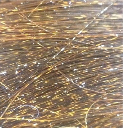
Fig. 7.2
Pili annulati. Characteristic alternating white bands on dermoscopy
Investigations Recommended
For Diagnosis
Diagnosis is based on hair shaft examination with a variety of methods (see Table 7.3).
Table 7.3
Evaluations for diagnosis of Pili Annulati
For diagnosis
Microscopic examination of hair shaft: the hair shaft presents alternating dark patches, which corresponds to air spaces in the cortex
Dermoscopy: air-filled cavities appear as white areas. Trichorrhexis nodosa in frequently present
Scanning electron microscopy: shows damaged cuticle and cortex structures adjacent to cavities within the hair shaft
For Treatment
No specific treatment exists. Gentle handling of the hair helps to decrease hair weathering
Woolly Hair
Clinical Features
Woolly hair resembles African hair. The hair is extremely curly, does not group in locks, and does not lie flat on the scalp [22]. The condition is rare and can be subdivided into three types based on clinical features and mode of inheritance (see Table 7.4).
Table 7.4
Subtypes of woolly hair
Type | Key characteristics |
|---|---|
Hereditary woolly hair (HWH) | Hair is normal in color |
Grows at normal rate, but may be short due to reduced duration of anagen phase | |
Autosomal dominaint inheritance | |
Familial woolly hair/woolly hair hypotrichosis (FWH/WHH) | Hair is lighter than unaffected family members |
Very short due to reduced anagen phase | |
Autosomal recessive inheritance | |
Woolly hair nevus (WHN) | Patch of abnormally curly hair, often lighter in color that surrounding hair |
Naxos syndrome (MIM 601214) | Woolly hair, palmoplantar keratoderma, and arrhythmogenic right ventricular cardiomyopathy |
Carvajal syndrome (MIM 605676) | Woolly hair, palmoplantar keratoderma, and dilated cardiomyopathy |
Hereditary woolly hair (HWH) is autosomal dominant inherited, and not associated with hair thinning or fragility [17]. The hair color is variable and the manageability improves with age.
Woolly hair is associated with palmoplantar keratoderma and cardiac abnormalities [23] in the Naxos syndrome (MIM 601214). The syndrome is autosomal recessive and caused by mutations in plakoglobin genes [24]. Mutations of desmoplakin cause Carvajal syndrome, which is characterized by woolly hair, palmoplantar keratoderma, and dilated cardiomyopathy.
Familial woolly hair (FWH) is autosomal recessive and associated with hair thinning. This condition, also known as woolly hair hypotrichosis, can be caused by mutations in either lipase H (LIPH) or lysophosphatidic acid receptor 6 (LPAR6) gene, encoding an LPA-producing enzyme PA-PLA1α and an LPA receptor LPA6, respectively. The hair is hypopigmented, sparse, thin, and short (Fig. 7.3) [25].
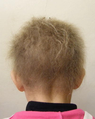
Fig. 7.3
Woolly hair hypotrichosis. Note sparse, short, blond wooly hair
Investigations Recommended
For Diagnosis
Diagnosis of woolly hair is based on clinical examination and the boiling water test. When immersed in boiling water, woolly hairs form regular spirals [28].
Microscopic examination of hair shaft is not useful for diagnosis.
On dermoscopy, woolly hair can demonstrate a short wavy “crawling snake” appearance [29].
Uncombable Hair Syndrome (Pili Trianguli et Canaliculi, Spun Glass Hair)
Clinical Features
Uncombable hair syndrome presents with blond, dry, and unruly hair that resists all efforts of styling. The overall appearance resembles synthetic doll hair. Eyebrows and eyelashes are normal and unaffected.
The condition usually appears during the first years of life and considerably improves with age when the hair becomes longer. Inheritance is known to be autosomal-dominant, but shows both complete and incomplete penetrance. The syndrome affects males and females equally. Besides the classic form, there is an acquired and partial form, where the abnormality is limited to the frontal or occipital area [31]. The condition is usually an isolated finding, but has been reported to occur with congenital abnormalities. Unruly hair similar to uncombable hair can be seen in Rapp-Hodgkin and loose anagen hair syndromes [32].
Investigations Recommended
For Diagnosis
Diagnosis requires dermoscopy or scanning electron microscopy [32]. The affected hair has a typical triangular or reniform-shape with longitudinal grooving and flattening.
Acquired Hair Shaft Disorders Without Fragility
Acquired Progressive Kinking of the Hair (APKH)
Clinical Features
APKH includes a variety of conditions (see Table 7.5) characterized by acquired curly, frizzy, and lusterless hair that resembles secondary sexual hair.
Table 7.5
Conditions described as acquired progressive kinking of the hair
Condition |
|---|
Whisker hair; kinking of the hair over preauricular areas of the scalp |
Acquired progressive kinking of androgen-dependent hair associated with thinning |
Rapidly progressive kinking of most or all the scalp hair without associated hair thinning |
Acquired reversible hair kinking before or after puberty |
Acquired hair kinking involving a localized non-androgen dependent area of the scalp |
Hair kinking after anagen/telogen effluvium (alopecia areata/drugs) |
Whisker hair is APKH over periauricular areas of the scalp. Whisker hair is short and curly and resembles hair of the beard. The condition is strongly associated with severe androgenetic alopecia [34]. Acquired progressive kinking of androgen-dependent hair represents a type of androgentic alopecia that is associated with poor prognosis in most cases [35].
Rapidly progressive kinking of most or all the scalp hair without associated hair thinning has been reported, albiet rare. APKH has been shown to be reversible in patients before and after puberty in certain instances [34, 36]. Acquired hair kinking can also be localized to non-androgen dependent areas of the scalp [37].
Investigations Recommended
For Diagnosis
APKH is a variation of AGA. Diagnosis can be confirmed principally with dermoscopy that shows more than 20 % variability or via scalp biopsy.
Microscopic Examination of Hair Shaft: Under light microscopy irregular twisting and periodic redution in hair shaft diamter can be observed.
Scanning Election Microscopy: Partial longitudinal grooves and longitudinal twisting of the hair shaft can be observed [41].
For Treatment
First Line Therapies
Since APKH is a variation of AGA, treatment options include finasteride 1 mg (E) [14] and topical minoxidil. However, minoxidil is not always effective.
Alopecia Areata (AA)
Clinical Features
Alopecia areata is a common non-cicatricial alopecia, characterized by patchy hair loss in the absence of inflammatory signs. The condition is an autommune T-cell mediated disorder, which leads to disruption of the normal hair cycle [42]. The disease affects both sexes at any age, but severe forms are more frequent in males, and often start during childhood.
The disease usually starts abruptly with one or multiple patches of hair loss that usually enlarge in a centrifugal way. Although AA frequently resolves spontaneously, relapses occur in a high percentage of patients. Diffuse shedding may or may not be present in the surrounding scalp. Peribulbar inflammation leads to interruption of the anagen phase with loss of dystrophic anagen roots [42]. AA may involve any hair-bearing skin, but is more common on the scalp (Fig. 7.4) and on bearded areas. Rarely, it may exclusively affect the eyelashes and/or the eyebrows [43].
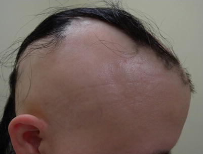
Fig. 7.4
Alopecia areata. Ophiasis pattern of hair loss
Severity of AA may be evaluated according to percentage of scalp involvement. Severe forms affect the entire scalp (AT, alopecia totalis) or all body hair (AU, alopecia universalis). Involvement of the scalp margin (ophiasis) is associated with a poor prognosis [44]. The affected scalp may be completely devoid of hair or may be covered by vellus, unpigmented, short hair. Acute AA is characterized by the presence of exclamation point and cadaverized hair. AA may also present with diffuse hair thinning without typical patches. The genetic inheritance of AA is polygenic. The condition affects first-degree relatives in about 20 % of cases, and monozygotic twins 42 % of the time [45, 46]. AA can be associated with a wide array of disorders, including various autoimmune diseases, nail abnormalities, and genetic syndromes (see Table 7.6).
Table 7.6
Associated disorders of alopecia areata
Associated disease | Findings |
|---|---|
Nail abnormalities | |
Superficial pitting with geometric pattern | Findings are especially prevalent in children |
Twenty-nail dystrophy (trachyonychia) | Nails are rough due to excessive longitudinal ridging |
Autoimmune disease | |
Thyroid autoimmune disease | Clinical/subclinical thyroiditis in up to 30 % of patients |
Celiac disease | Uncommon finding, but removing gluten does not change course of AA |
Vitiligo | 3–8 % of patients present with concurrent disease [47] |
Atopic disorders | |
Atopic dermatitis | Higher risk for development of AA; up to 31 % seen to develop [48] |
Genetic syndromes | |
Down syndrome | Prognosis with AA is unfavorable |
Polyglandular autoimmune syndrome type 1 | Rare condition; but reported in up to 33 % of patients [49] |
Investigations Recommended
For Diagnosis
Diagnosis in most cases is based on clinical examination by the presence of smooth discrete areas of hair loss and exclamation mark hairs.
Pull Test: The pull test is very positive if the disease is rapidly progressing.
Microscopic Examination: Microscopic examination shows telogen and dystrophic hair roots.
Dermoscopy: Dermatoscopic examination of the scalp may show a wide array of findings including exclamation mark hair, yellow dots, broken hairs, and black dots.
Histopathology: Is dependent on the chronicity of the disease. Acute AA has peribulbar lymphocytic infiltrates around anagen follicles described as “a swarm of bees.” Biopsy in chronic stage of AA shows follicular miniaturization, shift toward catagen and telogen follicles, vellus hair, and variability of inflammatory infiltrates [50].
For Treatment
Treatment of AA in children can be challenging and is based primarily on the use of topical agents in children under age 10.
In children over 10 years old, treatment is based on the same treatments as adults, which is based on extent of disease. Topical treatments and intralesional steroids are used in patients that have less than 50 % scalp involvement (Fig. 7.5).
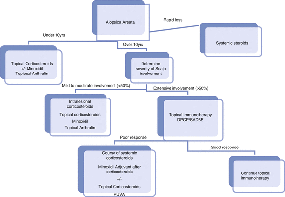
Fig. 7.5
Treatment algorithm for alopecia areata
In patients with more extensive scalp involvement, topical immunotherapies and oral corticosteroids can be considered [51].
In patients with extensive AA, wigs and tattooing of affected eyebrows can help mask the disease (see Table 7.7).
Table 7.7
Treatments for alopecia areata
First line treatment
Level of evidence
Triamcinolone acetonide 5–10 mg/ml scalp, 2.5–5 mg/ml beard/eyebrow monthly (10 years and up)
B
Clobetasol propionate 0.05 % applied under occlusive dressing 6 days per week for 3 months (preferred treatment over age of 14)
A
Clobetasol propionate 0.05 % foam (10 years and up)
A
Sensitization to DPCP or SADBE (preferred treatment for >50 % scalp involvement)
A
Second line treatment
Level of evidence
Topical anthralin; 0.5–1 %, short contact therapy (up to 2 h)
B
Topical PUVA 0.0001 % 8-MOP solution, three to four times weekly
B
Topical minoxidil 5 % foam or solution, daily to scalp
B
Third line treatment
Level of evidence
Oral prednisolone 200 mg once weekly for 3 months
B
Methylprednisolone iv 500 mg/day for 3 days a month for 3 months (10 mg/kg/day in children)
B
Oral prednisone 300 mg/month (5 mg/kg in children)
B
Oral dexamethasone 40 mg/month (5 mg/day for 2 consecutive days every week)
B
First Line Therapies
In patients who present with patchy alopecia or alopecia affecing the eyebrows, intralesional steroids are the preferred treatment (age >10 years). Triamcinolone acetonide should be diluted in saline solution at 5–10 mg/ml for injection in the scalp and 2.5–5 mg/ml for eyebrow and beard involvement. Maximum dosage should not exceed 40 mg per session. Treatement should be repeated every 4–6 weeks, but if no response is seen after 6 months, discontinue treatment and seek alternative treatment options [52].
In extensive disease (>50 % scalp involvement), topical immunotherapy with agents that induce allergic contact dermatits is preferred. These treatments have been shown to be superior to intralesional corticosteroids [53]. The two currently used sensitizers are diphenylcyclopropenone (DPCP) and squaric acid dibutyl ester (SADBE). Sensitization is obtained using a 2 % acetone SADBE or DPCP under closed patch test for 48 h. Treatment is performed with weekly application of the allergen diluted in acetone at a concentration that is able to induce mild scalp contact dermatitis. The concentration to achieve contact dermatitis can vary among different patients and even in the same patient during the treatment period. Acceptable regrowth (75 % or more) is seen in 38–63.8 % of patients [54, 55]. Moreover, hair regrowth can be achieved in 20 % of patients with alopeica totalis/universalis (AT/AU).
Clobetasol propionate 0.05 % applied under occlusive dressing nightly can improve hair growth significantly. However, in the authors’ experience this treatment is not a good option in children younger than 14 due to the risk of absorption and adrenal suppression [56]. Clobetasol propionate foam once a day has been shown to be effective in alopecia areata and can be utilized in children 10 years and older [57].
Second Line Therapies
Topical minoxidil 5 % solution or foam can be used in combination with topical or intralesional steroids. However, studies on efficacy of minoxidil are conflicting [58]. Minoxidil has been show to be useful to prevent relapses after interruption of systemic steroids [59].
Topical anthralin upto 1 % applied daily for 2 h or less can be used. Treatment induces mild erythema and irritation of the scalp. This treatment is an alternative option for children and mild forms of AA. However, effectiveness data is limited to case series [60].
Phototherapy can be used in the treatment of AA. Topical PUVA has been reported to regrow hair in 50–70 % of patients. However, studies are not RCTs and results have not been confirmed [61]. Topical PUVA using a 0.0001 % 8-MOP solution is applied using a soaked cotton towel at 37 °C for 20 min, followed by UVA irradiation three to four times a week, with cumulative UVA dose ranging between 60.9 and 178.2 J/cm2. Narrowband UVB is an option for patients with AT/AU, but efficacy rates are low, with 20 % responding to treatment [62]. Moreover, presence of hair limits UV penetration, and thus maintainance of hair regrowth is difficult.
Third Line Therapies
Systemic steroids can be used for acute severe AA. Various systemic steroid treatment protocols exists, but pulse steroids are perferred because of side effect profile (see Table 7.7). Studies have shown that pulse steroids are effective in acute progressive disease, but not in long-standing AT/AU. However, a high likelihood of relapse exists after discontinuation of systemic steroids [63]. Future promising treatments look at targeting the STAT1 pathway, including simvastatin and JAK-kinase inhibitors, which have shown promise in case reports and small studies [64, 65].
Loose Anagen Hair Syndrome (LAHS)
Clinical Features
LAHS is a benign, sporadic, or familial hair disorder that primarily affects children [66]. The condition is due to a defective anchorage of the hair shaft to the follicle, resulting in easily and painless pluckable hair. LAHS is more frequent in females than in males, occuring most commonly between the ages of 2 and 6. The typical patient is a young girl with short, blond hair that does not grow long. The condition usually improves spontaneously when the child grows up.
Three different varieties of LAHS have been catergorized by Olsen (see Table 7.8). Mutations in the gene encoding for the companion-layer keratin have been reported in some families with LAHS.
Table 7.8
Variants of loose anagen hair syndrome
Type | Major differences |
|---|---|
Type A | Characterised by decreased hair density |
Type B | Characterised by mainly unruly hair |
Type C | Characterised by increased hair shedding |
LAHS can cause diffuse thinning and irregular bald patches due to traumatic painless extraction of hair tufts. The hair is often dull, unruly, or matted (Fig. 7.6) [67]. LAHS is usually isolated, but may occur in association with hereditary or developmental disorders.
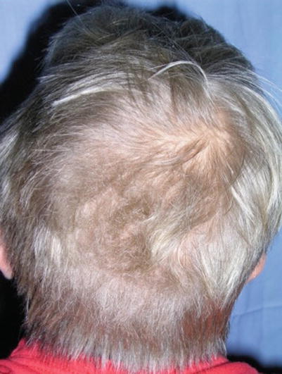
Fig. 7.6
Loose anagen hair syndrome. The hair is blond, short, and unruly
Investigations Recommended
For Diagnosis
Diagnosis is pricipably based on clinical features, pull test and trichogram.
Microscopic examination of hair shafts: LAH presents as anagen hair devoid of sheets; its bulb is often misshapen and its proximal portion often shows a ruffled cuticle.
Pull Test/Trichogram: Presence of LAH at pull test or trichogram may occur in controls. Thus, the diagnosis of LAHS should be made only if the trichogram shows at least 70 % LAH [68]. A negative pull test does not exclude the diagnosis.
Short Anagen Hair Syndrome
Clinical Features
This is a rare condition, which is characterized by a short hair cycle with an inability to grow long hair, and recurrent episodes of telogen effluvium [69]. The condition is usually diagnosed in childhood, and patients complain of short hair that does not grow and/or increased hair shedding (Fig. 7.7). The condition has also been reported in African Americans [70] and is usually sporadic [71]. However, familial cases have been reported, suggesting an autosomal dominant inheritance [69].
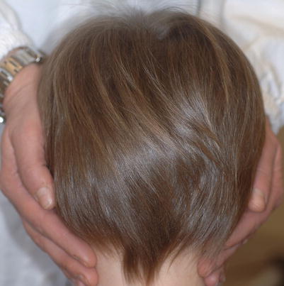
Fig. 7.7
Short anagen hair syndrome. The scalp hair is very short (less than 6 cm)
Investigations Recommended
For Diagnosis
The diagnosis is made by the characteristic clinical features and the finding of short (less than 6 cm long) telogen hair with a tipped point on pull test or trichogram [70].
The chief differential diagnosis is loose anagen syndrome.
For Treatment
Treatment for short anagen hair syndrome is not necessary because the condition usually improves after puberty, but increase in the hair length has been reported with minoxidil and cyclosporine [72].
Scarring Alopecia
Keratosis Follicularis Spinulosa Decalvans (MIM 308800)
Clinical Features
Keratosis follicularis spinulosa decalvans is a rare scarring alopecia that shows X-linked inheritance that has been mapped to Xp21-p23 [73]. This inherited condition usually becomes evident in infancy. The scalp presents with follicular keratotic papules and pustules that produces progressive cicatricial alopecia (Fig. 7.8a). Follicular papules are also evident on the eyebrows, eyelashes, and cheeks. The disease is slowly progressive and can produce severe alopecia.
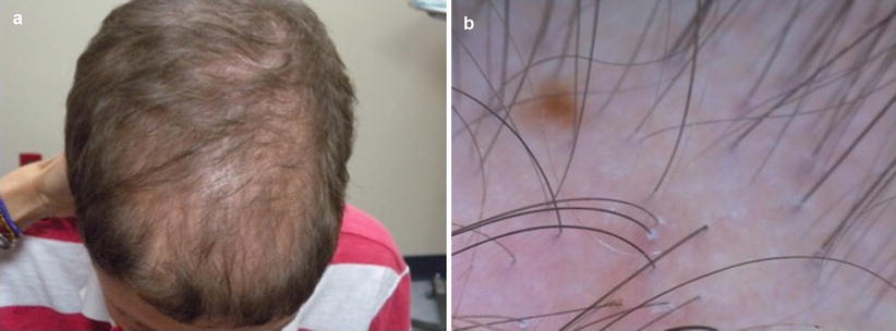
Fig. 7.8
(a, b) Keratosis follicularis spinulosa decalvans. (a) Scarring alopecia (b) Dermoscopy shows loss of follicular openings and peripilar casts
Investigations Recommended
For Diagnosis
The diagnosis is based primarily on characteristic clinical findings of scarring alopecia with papules and pustules.
Dermoscopy shows loss of follicular openings and peripilar casts (Fig. 7.8b). Findings are similar to those observed in lichen planopilaris.
For Treatment
No specific treatment exists. All treatments are minimally effective and the disease is slowly progressive.
Emollients, topical steroids and keratolytic agents can provide symptomatic relief.
Case reports reported improvement with oral retinoids, dapsone, and tetracyclines.
Eruptive Vellus Hair Cysts (EVHC)
Clinical Features
Eruptive vellus hair cysts are rare benign neoplasms that present as multiple smooth or umbilicated papules, predominately on the chest and abdomen [74]. They primarily occur in children and young adults, and have been noted to occur concurrently with steatocystoma multiplex. Most cases are sporadic, but an autosomal dominant pattern has been reported [75]. EVHC has been associated with paronychia congenita, trichostasis spinulosa, hidrotic ectodermal dysplasia, and anhidrotic ectodermal dysplasia [76].
Investigations Recommended
For Diagnosis
Although clinical examination can provide clues to the diagnosis, definitive diagnosis is made with biopsy or incision into top of the lesion, followed by expression of cystic material.
Dermoscopy: On Dermoscopy, EVHCs have well-defined white to yellow round structures with erythematous brownish halos. A central gray/blue color can be seen that corresponds to the melanin in the hair shaft of the cyst [77]. Dermoscopy is helpful because it can aide in differentiation of EVHCs from molluscum contagiosum, which has a polylobular white to yellow amorphous center with a crown of hairpin vessels at the periphery [75].
Histopathology: EVHC have small epidermoid cysts, which contain multiple vellus hairs.
For Treatment
EVHC present a cosmetic problem and can be bothersome when occurring on the face. A variety of medical and surgical treatment options have been used with variable success (Table 7.9). However, 25 % of lesions spontaneously regress without treatment.
Table 7.9
Treatments for eruptive vellus hair cyst
Treatment
Related posts:Stay updated, free articles. Join our Telegram channel
Full access? Get Clinical Tree
 Get Clinical Tree app for offline access

|
|---|