For diagnosis
Clinical–facial eruption of pustules that begins at age 5–30 days. It resembles neonatal acne but lacks comedones and is associated with colonization with Malassezia
Giemsa stain–yeast form, neutrophils
For treatment
None, self-limiting
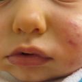
Neonatal/Infantile Acne (Fig. 2.1)
For diagnosis |
Clinical–comedones and inflammatory papules and/or comedones. It usually begins in the first year of life and is secondary to a physiologic increase in adrenal and gonadal androgens. |
If severe consider work-up for hyperandrogenism |
For treatment |
Treat to avoid formation of pitted scarring from acne |
For Severe Acne Consider
Oral erythromycin, azithromycin, or trimethoprim-sulfamethoxazole | D |
Oral isotretinoin | D |
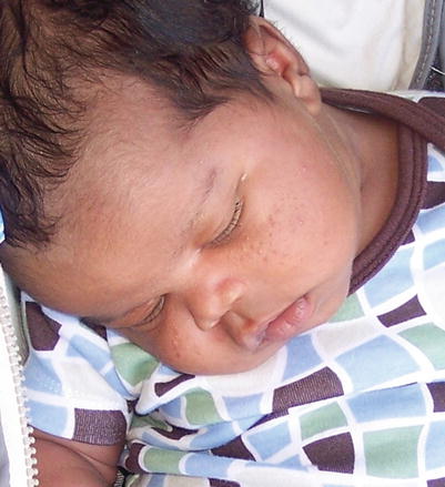
Fig. 2.1
Neonatal acne
Seborrheic Dermatitis
For diagnosis |
Clinical–greasy yellow scale and erythematous patches on the face, scalp, ears, and intertriginous areas. Appears between 2 and 10 weeks of age and may be associated with colonization of Malassezia furfur |
Consider culture for bacteria and candida for any weeping intertriginous area |
KOH preparation and fungal culture to exclude superficial dermatophyte infection |
For treatment |
None, self-limiting within a few weeks to months |
Emollients. For thick scale, application of mineral or baby oil followed by gentle scalp massage with a soft toothbrush | A |
Frequent shampooing with gentle shampoo or anti-seborrheic shampoo | E |
Bifonazole 1 % shampoo | B |
Ketoconazole 2 % shampoo | A |
Ketoconazole 2 % cream | B |
Hydrocortisone 1 % cream | B |
Acrodermatitis Enteropathica
For diagnosis |
Clinical–well demarcated scaly perioral and acral plaques, which may be accompanied by alopecia and diarrhea. It is secondary to an autosomal recessive mutation in intestinal zinc-specific transporter gene SLC39A4. Acquired forms of zinc deficiency will have the same clinical presentation and may be secondary to inadequate intake, excessive losses, malabsorption, and increased demands. Mothers may have low zinc secretion into milk caused by a mutation in the SLC30A2 gene, which encodes the transporter responsible for secreting zinc into breast milk |
Blood plasma or serum zinc levels, fasting; serum zinc levels <50 μg/dl |
Low alkaline phosphatase levels |
Urine zinc excretion |
Physical exam and routine laboratory evaluation, lipid profile, copper levels |
Genetic testing |
For treatment |
Blood plasma or serum zinc levels q 3–6 months |
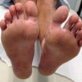
Acropustulosis of Infancy
For diagnosis |
Clinical–pruritic recurrent pustules on palms and soles |
Rule out scabies |
Skin biopsy–intraepidermal pustules with neutrophils, occasional eosinophils |
For treatment |
None |
Potent topical corticosteroids | D |
Oral antihistamines | E |
Aplasia Cutis Congenita [14] (Figs. 2.2 and 2.3)
For diagnosis |
Clinical–discrete ovoid defect covered with a membrane that may be bullous or flat at birth that eventually heals with a scar. Aplasia cutis congenita may present sporadically or may be inherited as part of a syndrome |
Thorough history and physical for other developmental anomalies |
If hair collar sign (ring of longer, darker hair around the defect) and midline, need evaluation for underlying neural tube defect; ultrasound or MRI if concerned |
For treatment |
Follow-up proper formation of scar |
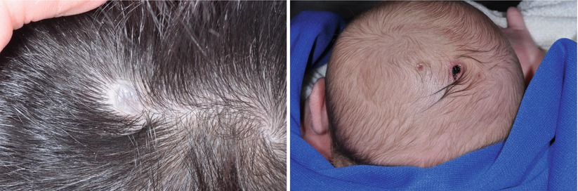
Figs. 2.2 and 2.3
Aplasia cutis congenita
Bronze Baby Syndrome
For diagnosis |
Clinical–hyperpigmentation of the skin, serum, and urine during treatment for neonatal jaundice with phototherapy. It occurs in infants with cholestasis and elevated levels of unconjugated and conjugated bilirubin |
Evaluate for underlying cause of jaundice |
Evaluate for underlying hepatocellular disease |
For treatment |
Monitor for jaundice, cholestasis, hepatocellular disease |
Congenital/Neonatal Herpes Simplex Virus (HSV)
For diagnosis |
Clinical–vesicles on an erythematous base; three recognized syndromes: skin, eyes, and mouth infection (SEM); disseminated infection; central nervous system infection |
Viral Culture (swab from mouth, nasopharynx, conjunctiva, anus, and any vesicles), Viral DFA or immune peroxidase slide test, PCR (skin vesicle, CSF, blood), Tzanck Preparation, ALT elevation, skin biopsy |
Must rule out CNS disease |
CT brain, MRI brain, EEG |
For treatment with acyclovir |
Serial absolute neutrophil count (ANC) twice weekly |
Adjust dose of Acyclovir for renal failure or sustained ANC <500 mm3 |
For infants with CNS disease- consider daily suppressive therapy with oral acyclovir for 6 months after parenteral regimen |
Congenital/Neonatal Candidiasis
For diagnosis |
Clinical; congenital–pustules on palms and soles birth to first few days of life, may have respiratory distress; neonatal–diaper (beefy erythema and satellite pustules or vesicles), oral thrush (white plaques on oral mucosa), and also commonly associated with intertrigo (erythema and maceration in skin folds). This warrants a high index of suspicion in premature and immunocompromised infants |
Smear of pustule with KOH, Giemsa, Gram, or calcofluor stain (budding yeast or pseudohyphae), fungal culture |
PCR, restriction fragment endonuclease digestion of chromosomal DNA, electrophoretic karyotyping, Southern blot hybridization analysis with DNA probes, B-glucan assay, gas chromatography mass spectrometry for D-arabinitol, buffy coat smear microscopy |
CBC (leukocytosis), Glucose (elevated) |
Congenital–evaluate placenta and umbilical cord for lesions; if suspect disseminated systemic disease (premature and low birth-weight), must culture blood, urine, cerebrospinal fluid |
For treatment |
Monitor for sepsis |
Follow monitoring guidelines for any PO or parenteral antifungals |
Thrush–nystatin solution (100,000 u/ml) applied to oral mucosa 4×/day; fluconazole (2–3 mg/kg/day) | A |
For localized disease–topical anti-yeast preparations, Imidazoles, nystatin, allylamines | A |
Invasive–oral fluconazole, itraconazole, amphotericin | A, B (amphotericin) |
Prophylaxis in infants <1 kg-Fluconazole IV | A |
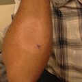
Staphylococcus Aureus Pustulosis (Fig. 2.4)
For diagnosis |
Clinical–discrete vesicles and pustules, or superficial erosions and crust |
Bacterial culture and gram stain of fluid from vesicle, pustule, or beneath crust. Nasal swabs to evaluate for S. Aureus carriage |
Consider work-up for deeper or systemic infection if any constitutional signs of illness (fever, temperature instability, irritability, lethargy, etc.) |
For treatment |
Monitor for signs of deeper or systemic infection |
Monitor sensitivities of culture |
Consult with colleagues and local resources regarding resistance patterns in your hospital and community |
Oral antibiotics–choose agent based on sensitivity and local resistance patterns | A |
Topical antibiotic ointment (mupirocin, fusidic acid, or retapamulin ointments) | A |
Decolonization of neonates and close contacts | A (adults– trial results pending in neonates) |
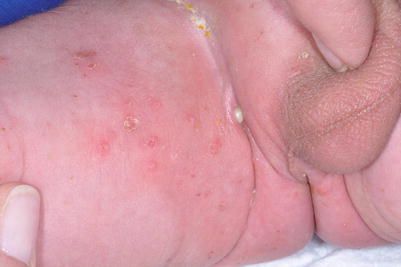
Fig. 2.4
Staphylococcal pustulosis
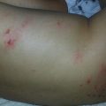
Neonatal Scabies (Fig. 2.5)
For diagnosis |
Clinical–pruritic contagious infestation of the Sarcoptes scabiei mite that commonly involves the palms, soles, and axillae but also may involve the scalp of infants. Burrows, vesicles, erythematous papules, and nodules may be present |
Mineral oil examination–apply a drop of mineral oil and scrape with No. 15 blade then smear contents onto glass slide, cover with mineral oil and evaluate for mite, eggs, or feces |
Dermoscopy |
For treatment |
May have pruritus for 1 month following treatment |
Permethrin 5 % cream–approved for over 2 months of age but commonly used under 2 months of age, traditionally applied to all skin from neck down and rinsed after 8 h, but scalp must also be treated in infants; should be repeated after 1 week | A |
Sulfur 6 % ointment–safe in infants and pregnant women; apply as above for 3 consecutive nights and rinse 24 h after last application | D |
Treat all close contacts | E |
Launder all linens following treatment
Related posts:Stay updated, free articles. Join our Telegram channel
Full access? Get Clinical Tree
 Get Clinical Tree app for offline access

|