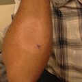
For diagnosis
Clinical examination for classic-appearing burrows, often with a vesicle or small pustule at the end of a burrow
Skin scraping with mineral oil for microscopic identification of mites, ova (eggs) or scybala (scabies feces)
Biopsy if diagnosis is uncertain
High-magnification videodermatoscopy or PCR of scale can confirm diagnosis, though very rarely necessarily
Management Strategies
Treat all close contacts to limit spread of disease |
Wash clothing, sheets and towels in hot water (140 °F, 60 °C) and dry with high heat [3] |
Vacuum and clean living quarters |
Put stuffed animals, other toys and fomites in a plastic bag for 72 h |
Of note, pets do not need to be treated since they cannot harbor these mites |
Therapies [3–14]
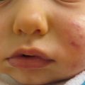
In general, the face and scalp do not need to be treated unless the patient is an infant or immunocompromised. Resistance to topical therapies has begun to emerge.
Table 19.1
First line therapies
Permethrin 5 % cream, applied from the neck down and rinsed off after 8–14 h, repeat in 1 week | A |
Contraindicated in infants under 2 months of age | |
In pregnant women, shorten the application to 2 h prior to rinsing off | |
Ivermectin 200 μg/kg per dose, taken orally for one dose; repeat in 2 weeks: | A |
Off-label use | |
Consider in severe cases, crusted scabies or immunocompromised individuals | |
Not recommended in children under 15 kg of weight or in pregnant or nursing women | |
Malathion 0.5 % lotion to dry hair for 8–12 h then rinsed off; repeat in 7–9 days: | C |
FDA approved over 6 years of age | |
Flammable |
Table 19.2
Second line therapies
Sulfur 6–10 % ointment compounded into petrolatum or mineral oil applied from the neck down 1–2 times daily for 3–5 days and rinsed off 24–48 h after the final application | B |
Can be used in pregnant women and infants | |
Ivermectin 1 % lotion to applied at a dose of 400 μg/kg, repeated in 1 week | A |
Crotamiton 10 % cream applied from the neck down 1–2 times daily for 5 consecutive days and rinsed off 24 h after the final application; repeat one overnight application 1 week after initial day of application: | B |
Resistance common | |
Intralesional corticosteroids for nodular lesions | E |
Topical steroids and antihistamines for post-scabietic pruritus | E |
Table 19.3
Third line therapies
Benzyl benzoate 25 % lotion applied from the neck down every other night for 3 applications: | B |
Should not use in infants and young children | |
Not available in the United States | |
Lindane 1 % lotion applied from the neck down and rinsed off after 8–12 h neck, repeat in 1 week: | A |
Avoid under 2 years of age as well as pregnant or nursing women given possible neurotoxicity | |
Banned in California | |
Resistance common | |
American Academy of Pediatrics does not recommend its use in children | |
Monosulfiram soap | C* |
Topical thiabendazole solution | B* |
Co-trimoxazole | D* |
Natural pyrethrins | C |
Topical dimethicones | A |
Chiggers (Harvest Mites, Jiggers, “RED BUGS”)
Clinical Features
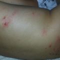
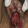
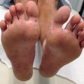
Chiggers, Trombicula alfreddugesi, are unique in that only the six-legged larvae, and not the eight-legged adults or nymphs, affect humans and animals. Found most commonly in the southern United States, the larvae are found in vegetation, and attaches to those who pass by. Chigger infestation is self-limited, usually seen in the summer and fall, and presents as intensely pruritic, grouped, edematous, erythematous 1–2 mm papules or papulovesicles, with a hemorrhagic punctum at sites of constricted clothing (elastic in socks, edges of underwear, belts at the waist) [15]. In boys and men, chiggers can also be found on the penis, causing “summer penile syndrome” with symptoms of penile swelling, pruritus, and dysuria. The larvae inject an irritating substance into the skin, causing the pruritus, before falling off the skin or being scratched off. Secondary infection may develop.
Investigations Recommended
Investigations recommended |
None |
Management Strategies
Permethrin on clothing for prevention |
Washing immediately after exposure may help prevent symptoms |
Therapies [16]
Supportive care with antipruritics (camphor, menthol), cool compresses, topical anesthetics (pramoxine), topical or intralesional corticosteroids, and oral antihistamines | E* |
Vinegar (5 % acetic acid) may help with post-exposure prophylaxis and symptomatic relief of pruritus | E* |
Cheyletiella (Walking Dandruff)
Cheyletiella are relatively larger non-burrowing mites (approximately 0.4 mm in length) caused by animal-specific mites. They are found on dogs, cats, and rabbits and are caused by Cheyletiella yasguri, Cheyletiella blakei, and Cheyletiella parasitivorax, respectively. Affected animals have fine, flaking scale, but may not exhibit any symptoms of infestation. These mites do not live on human skin; they quickly bite a nearby human and return to their animal hosts, causing grouped pruritic papules, vesicles, bullae, and wheals.
Investigations Recommended
For diagnosis |
Have pets evaluated by a knowledgeable veterinarian |
Brushings from animal’s hair and place in a plastic bag with alcohol where mites will float while hair and scales sink |
For treatment |
None |
Management Strategies
Management strategies |
None |
Table 19.4
Therapies
Supportive care | E* |
Dips and shampoos from veterinarians for pets, including fipronil, permethrin, and amitraz | E* |
Grain and Avian Mites
Clinical Features
As the name suggests, grain mites are found in grain and straw. Bites from these mites result in intensely pruritic macules, papules, vesicles, or pustules followed by urticarial wheals without burrows. Fever and purpura may develop in more severe cases. Avian mites (also known as fowl mites or bird mites) are rarely found on humans, but instead are found in nearby birds, nests, clothing, and bedding. Two different genera of avian mites cause accidental skin eruptions, termed gamasoidosis in humans: Dermanyssus and Ornithonyssus. Eruptions are typically a widespread pruritic papular dermatitis with a morbilliform appearance, with possible vesicles and urticarial wheals without burrows. Eruptions from either type of mite are self-limited.
Investigations Recommended
For diagnosis |
Search for avian mites on clothing, bedding, nests and nearby birds |
Management Strategies
Management strategies |
None |
Table 19.5
Therapies
Supportive care | E* |
Demodex
Clinical Features
Demodex mites, Demodex folliculorum and Demodex brevis, are normally found in human pilosebaceous units of the head and neck. In some susceptible individuals, they may cause a pruritic folliculitis on the face and/or trunk comprised of both papular and pustular lesions, occasionally with a perioral affinity resembling rosacea. Demodex often may affect the eye as well, leading to blepharitis, chalazions, and other findings. This eruption is more common in adults and, in children, may suggest immunocompromise.
Investigations Recommended
For diagnosis |
A skin biopsy may be helpful when diagnosis is uncertain |
Management Strategies
Management strategies |
None |
Permethrin 5 % cream applied to affected area, wash off in 8–14 h, and repeat in 1 week if needed | E* |
Crotamiton 10 % to affected areas twice daily until improved | E* |
Benzoyl peroxide 2–10 % wash or gel to affected area daily until improved | E* |
Oral metronidazole 15 mg/kg/day or 750 mg/day in divided doses for 2–3 weeks | E* |
Oral ivermectin 200 μg/kg per dose, taken orally for one dose; can be taken in conjunction with oral metronidazole | B* |
Topical ivermectin 1 % cream daily for 12 weeks, metronidazole 0.75 % or 1 % gel or cream, sulfur, sodium sulfacetamide, benzoyl benzoate, lindane, hexachlorocyclohexane or camphor oil have also been reported | E* |
Ticks
Clinical Features
Ticks are round arachnids with specialized mouth parts for sucking blood. There are three categories of ticks: hard ticks (Ixodidae), soft ticks (Argasidae), and Nuttalliellidae. Only hard and soft ticks bite humans; however, hard ticks transmit the majority of diseases because they can remain attached to human skin, whereas soft ticks do not. Most tick bites are painless, and transmission of diseases usually result only after 24 h of tick attachment. Tick-borne diseases often have specific vectors, and more than one disease can be transmitted at a time. Ixodes scapularis and I. pacificus transmit Lyme disease, babesiosis, human granulocytic ehrlichiosis (human anaplasmosis), and in Europe, may cause viral encephalitis. The Dermacenter ticks, D. variabilis (the American dog tick) and D. andersoni (the Rocky Mountain wood tick), are major vectors for Rocky Mountain Spotted Fever in the eastern and western United States, respectively. D. andersoni also transmits Colorado tick fever, Q fever, and tularemia. Amblyomma americanum (the lone star tick) is a vector for human monocytic ehrlichiosis. Ornithodoros soft ticks transmit borrelial relapsing fever. Ixodes ticks tend to bite the torso; Amblyomma ticks, the lower legs, buttocks and groin; Dermacentor ticks, the head, neck, and upper trunk.
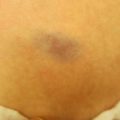
Patients may present with the offending tick still attached, often thinking that it is a new mole, as the attached tick slowly engorges itself with blood before falling off up to 2 weeks later. Hypersensitivity reactions to bites may cause erythematous papular, nodular, bullous, and ulceronecrotic lesions that are usually pruritic. A classic sign of tick bites is the “comet” sign, where many bites spread from initial points in the distal areas of the ankles and legs [23].
Tick bites may lead to secondary infection, alopecia, cutaneous lymphoid hyperplasia, and granulomas (possibly related to retained mouthparts). “Tick bite pyrexia” may develop in some patients, causing fever, chills, vomiting, headache, flu-like illness, and abdominal pain. Systemic symptoms usually resolve within 36 h of tick removal. Notably, Dermacentor ticks hidden in the scalp may potentially cause life-threatening tick paralysis. This condition is more common in children, and resembles Guillain-Barré syndrome with a reversible ascending flaccid paralysis. The mortality rate is 10 % due to respiratory failure. Removal of tick parts leads to rapid resolution.
Investigations Recommended
For diagnosis |
Thorough clinical examination for evidence of tick bites, attached ticks, or residual tick parts; if found, removal of entire tick including mouth parts and attachment cement, and without squeezing its abdomen |
Lyme serology when appropriate |
Rickettsial immunofluorescence and immunoperoxidase studies when appropriate |
ELISA, PCR, and immunofluorescence available for some viruses such as West Nile |
Management Strategies
Clothing to try to prevent bites: |
Light-colored and long-sleeved clothing |
Close-toed shoes |
Tuck pants into socks |
Insect repellents: |
DEET or picaridin to exposed skin |
Permethrin applied to clothing |
Control of tick populations is important: |
Exclusion of animal hosts |
Removal of leaf debris |
Treatment of ticks on animal populations and pets |
Table 19.7
Therapies
Supportive care | E* |
Potent topical or Intralesional corticosteroids for the tick bite reaction, if needed | E* |
Diseases are treated specifically and beyond the scope of this chapter, though most tick-borne diseases such as Rocky Mountain Spotted Fever respond to doxycycline, which is the first-line therapy even in children and pregnant women; in pregnant women, if the disease appears to be mild, chloramphenicol may be an alternative choice of therapy | |
Order Araneida
Spiders are found worldwide and play an important role in controlling insect populations. Spiders tend to bite out of self-defense and bites are common, though the offending spider is usually not identified. The two most clinically relevant species in the United States are the black widow (Latrodectus mactans) and brown recluse (Loxosceles reclusa) species and will be discussed here.
Black Widow Spider (Latrodectus Mactans)
Clinical Features
The black widow spider is a black- or brown-colored spider with a reddish hourglass visible on the ventral abdomen, and is found throughout the North American continent and Cuba. It has a potent depolarizing neurotoxin, alpha-latrotoxin, and female spiders are more dangerous due to their larger size. A black widow spins webs in cool, dark places and tends to bite only if threatened. Bites appear as two red marks with surrounding edema, and are normally seen on exposed skin, buttocks, or genitalia. The bites result in severe pain within minutes, and over the course of hours, chills, vomiting, muscle cramps, abdominal rigidity, and partial paralysis may ensue. A morbilliform eruption may sometimes be present, and several reports of resulting priapism exist in the literature [24]. Children may develop profuse sweating and become more agitated and irritable. Most black widow bites run a self-limited course, though a small minority of bites may be lethal.
Investigations Recommended
For diagnosis |
Identification of the offending spider, when possible |
Management Strategies
Management strategies |
Medical observation if any systemic symptoms develop |
Specific antivenin, given up to 90 h after the bite | A* |
Pain control, possibly requiring intravenous opiates | E* |
Benzodiazepines (diazepam) as needed, though they do not shorten duration of symptoms and may have side effects | E* |
Calcium gluconate as needed for associated tetany | E* |
Antibiotics if secondarily infected | E* |
Brown Recluse Spiders (Loxosceles reclusa)
Clinical Features
Many species of Loxosceles spiders can be found throughout the world. Loxosceles reclusa, the brown recluse spider, is found hidden in dark, dusty areas, such as attics and basements, predominantly in the Southeastern, Midwestern, and Southwestern United States. It is a brown spider with a dark-brown, violin-shaped mark on the dorsal cephalothorax measuring around 1 cm in size. The primary toxic enzyme is sphingomyelinase D, which has numerous hematologic effects, including red blood cell lysis and skin necrosis due to neutrophil activation. Other enzymes found include alkaline phosphatase, esterase, ATPase, and hyaluronidase. The spider tends to bite only in self-defense, and these slow-healing bites are usually found on the extremities. Mild cases result in a self-resolving urticarial reaction. Classically, a bite causes localized pain, pruritus and erythema within 6 h and progressively becomes more painful, ulcerated, and necrotic over the subsequent 18 h. Lymphangitis and gangrene may develop over the following week, occasionally together with a generalized petechial or morbilliform eruption, as well as fever, chills, nausea, and arthralgias. Serious systemic findings are rare, but may include necrotizing fasciitis, severe hemolysis, renal insufficiency, pulmonary edema, disseminated intravascular coagulation, and shock.
Investigations Recommended
For diagnosis |
Identification of the offending spider, when possible |
Complete blood count and fibrin split products |
Management Strategies
Management strategies |
Seek medical attention unless mild symptoms only |
Table 19.9
First line therapies
Rest, ice, and elevation of bite site | E* |
Tetanus prophylaxis, if needed | E* |
Antivenin, if available | E* |
Conservative debridement if necrotic and no longer spreading | E* |
Table 19.10
Second line therapies
Sulfone (dapsone) 100–300 mg PO daily | E* |
Antibiotics if secondarily infected | E* |
Aspirin, antihistamines (cyproheptadine) and tetanus vaccination can be considered | E* |
Intravenous fluids and systemic corticosteroids (1–2 mg/kg/day) if severe skin lesions, systemic symptoms and in small children, though controversial | E |
Order Scorpionida
Scorpions
Clinical Features
Scorpions are nocturnal, tropical arachnids with a stinger containing venom at the end of a curved, elongated tail. They are found worldwide, and in the United States, Centruroides exilicauda is the most common species, found most commonly in the southern states. Scorpions tend to hide in dark places and sandboxes, and sting only by accident or in self-defense. Stings deposit two toxins, a hemolytic toxin and a neurotoxin, typically leading to pain and paresthesias often out of proportion to the erythema and edema seen, accompanied by tachycardia and hypertension. Locally, other cutaneous findings that may be seen include petechiae, purpura, bullae, necrosis, and lymphangitis. The deposited neurotoxin may potentially cause salivation, vomiting, colicky abdominal cramps, psychomotor agitation, convulsions, cardiac arrhythmias, acute pulmonary edema [23] and, rarely, respiratory paralysis and death. In general, children are at higher risk for severe envenomation, as most fatalities from scorpion stings are reported in children younger than 10 years old [31, 32]. Notably, stings from the Egyptian scorpion lead to death from respiratory failure in 50 % of children.
Investigations Recommended
Investigations recommended |
None |
Management Strategies
Management strategies |
Observe children after stings closely for at least 4 h, given higher risk of severe reaction to venom |
Apply a tourniquet proximal to the area of sting, when possible, though controversial | E* |
Local anesthetics (1–2 mL lidocaine 2 % or bupivacaine 0.5 %) without vasoconstrictors repeated every 30–60 min for up to 3 injections or application of ice to the bite | E* |
Anti-venom, if available, best given within 4 h | B |
If severe, admission to the Intensive Care Unit: intravenous fluids, sedation (intravenous or oral midazolam 0.05–2 mg/kg), anti-arrhythmics, anti-hypertensives (prazosin 30 μg/kg orally every 6 h for 48 h or until clinical improvement), and calcium-channel blockers as needed | E |
Intravenous corticosteroids no longer recommended | E |
Class Insecta
Insects have three body parts—a head, thorax and abdomen—and have three pairs of legs. This class of bugs is very clinically important, since approximately 25 % of reports of anaphylaxis are due to insect stings. Often times, only one family member may be bitten, even when all are exposed to the same environment.
Order Phthiraptera, Suborder Anoplura
Lice (or louse, if singular) are wingless, `flat-bodied insects that have been infesting humans worldwide for thousands of years. Three types of lice are clinically important—head lice (Pediculus humanus var. capitis), body lice (Pediculus humanus var. corporis), and pubic lice, also known as head crab or crab lice (Phthirus pubis). Lice are obligated to live off human blood, and measure 1–4 mm in size. Head and body lice are elongated and similar in appearance, whereas crab lice resemble a miniature version of the food.
Pediculosis Capitis (Head Lice)
Clinical Features
In children, head lice cause the most common of the lice infestations, and are most frequently seen in children ages 3–12 and their parents. It affects all races, though African-American children are less often infected. Head lice usually cannot survive away from the human host more than 36 h. Adult lice are approximately 1–3 mm in size and feed on the scalp every 4–6 h. Females live for approximately 1 month and lay five to ten eggs daily. Eggs, called nits, are 0.8 mm in size, and viable nits are usually found cemented by a proteinaceous matrix to the hair within 0.6 mm of the scalp, but can be farther away in warmer climates. Nits develop into adult lice over the course of 2 weeks. Bites from head lice are painless, and the hallmark of disease, intense scalp pruritus, usually manifests within 2–6 weeks of infestation. The diagnosis is made by identifying the louse or nits in the scalp. Erythematous macules, papules, excoriations, scaliness or secondary bacterial infection can sometimes be seen on the scalp. Cervical or suboccipital lymphadenopathy may also be present.
Investigations Recommended
For diagnosis |
Thorough clinical examination for lice and nits |
Mount on a glass slide for microscopic evaluation if needed |
Dermoscopy may be of benefit |
Management Strategies
Screen all close contacts and treat those found to have lice or nits simultaneously; empirically treat if any close contacts share a bed or comb |
Clean hats and headgear |
Vacuum living quarters |
Over-the-counter permethrin 0.5 % spray for fomites (furniture, etc.) |
Manual removal of nits with a fine-toothed nit-removal comb on damp hair; vinegar may help to loosen the cement |
The American Academy of Pediatrics recommends against no-nit policies since they have not been found to be effective [40] |
Wash clothing, sheets, and towels hot water (at least 122 °F, 50 °C) or on high heat in the dryer for a minimum of 10 min; place items not appropriate for laundering in a plastic bag for 1–2 weeks |
Therapies
Of note, there is increasing resistance to some of these therapies, particularly permethrin and lindane. No therapies are reliably ovicidal, so repeat treatment in 1 week is recommended for all of the following. This also helps to fight growing resistance, which has already been reported with permethrin, pyrethrins, lindane, and malathion. Until safety is established for the following therapies, only mechanical removal of lice and nits is recommended for children under 2 years old, except for permethrin 1 % cream, which is FDA approved down to 2 months of age [3, 4, 13, 41–53].
Table 19.13
First line therapies
Over-the-counter permethrin 1 % lotion or cream rinse to damp hair and rinse off in 10 min without subsequent shampooing of hair for at least 24 h | A |
Permethrin 5 % cream applied to hair and scalp overnight for 8–14 h and rinsed off in the morning | E* |
Pyrethrins 0.3 % +/− piperonyl butoxide 4 % shampoo or mouse applied for 10 min then rinsed off: | A |
Avoid in patients allergic to chrysanthemums and related plants such as ragweed |
Table 19.14
Second line therapies
Malathion 0.5 % lotion to dry hair for 30 min to 12 h then rinsed off; repeat in 7–9 days: | A |
FDA approved over 6 years of age | |
Flammable | |
Spinosad 0.5–1 % topical suspension applied to dry hair for 10 min then rinsed off; repeat in 7 days: | A |
Do not use in neonates and in lactating women | |
Benzoyl alcohol 5 % lotion to dry hair for 10 min then rinsed off: | A |
FDA approved over 6 months of age | |
Can be used in pregnant or lactating women | |
Ivermectin 200–400 μg/kg per dose, taken orally for one dose: | A |
Off-label use and not recommended in children under 15 kg of weight or in pregnant or nursing women | |
Ivermectin 0.5–1 % lotion to dry hair and scalp and rinse out with water in 10 min applied only once | A |
Crotamiton 10 % lotion twice daily for 5 days | E |
Topical occlusion therapy with petroleum jelly, dimethicones, or other occlusive agents | A |
Mechanical removal (wet combing) of lice and nits every 3 days for 2 weeks | A |
Table 19.15
Third line therapies
Lindane 1 % shampoo or lotion to dry hair for 4 min (shampoo) or overnight (lotion) then rinsed off; applied only once and not repeated: | A |
Avoid under 2 years of age as well as pregnant or nursing women given possible neurotoxicity | |
Banned in California | |
American Academy of Pediatrics does not recommend its use in children | |
Carbaryl 0.5 % lotion or shampoo for 8–12 h then rinsed off: | B |
Not approved in the United States | |
1,2-octanediol 1 % spray twice weekly to washed and dried hair for 6 weeks | A |
Tocopheryl acetate 20 % spray to dry hair for 20 min, then washed | A |
Oral trimethoprim-sulfamethoxazole 8–10 mg/kg/day divided BID for 10 days (alone or together with permethrin 1 %): | B |
Off-label and mixed data | |
Alternative considerations though not studied well: | E |
Greasy occlusion of the scalp with Cetaphil Gentle Skin Cleanser, petrolatum jelly, styling gels, and various oils | |
Shaving the head | |
Hot air to the scalp for 30 min | |
Herbal shampoos |
Pediculosis Corporis
Clinical Features
The body louse measures 2.4–4 mm in length and is most often found in areas of poor hygiene, poverty, and overcrowding. Unlike the other two types of lice, body lice are vectors of several human diseases, including epidemic typhus (Ricketssia prowazekii), relapsing fever (Borrelia recurrentis), trench fever, and bacillary angiomatosis (Bartonella quintana). The louse is rarely found on human skin, but rather, resides in the seams of clothing, where its nits can also be found, and it survives for up to 1 month away from the host. Bites from the body louse are usually painless, and result in erythematous or copper-colored macules, pinpoint papules or wheals, with a hemorrhagic punctum. Excoriations, secondary bacterial infections, furuncles, and lymphadenopathy may be found. Unlike scabies, the hands and feet are not typically involved.
Investigations Recommended
For diagnosis |
Examine clothing for identification of body louse |
Management Strategies
Wash clothing in hot water (at least 122 °F, 50 °C) or on high heat in the dryer for 30 min, and iron all seams |
Treat clothing and fomites with permethrin 0.5 % spray, malathion 1 % powder, or dusting powders containing DDT |
Abandon infested mattresses and other fomites for 1 month, or discard |
Treat all household and close contacts |
Improve personal hygiene as able |
Table 19.16
First line therapies
Permethrin 5 % cream applied from the neck down and rinsed off after 8–14 h, repeat in 1 week: | E* |
Contraindicated in infants under 2 months of age | |
Pyrethrins 0.3 % +/− piperonyl butoxide 4 % shampoo or mousse applied for 10 min then rinsed off: | E* |
Avoid in patients allergic to chrysanthemums and related plants such as ragweed |
Table 19.17


Second line therapies
Related posts:
Stay updated, free articles. Join our Telegram channel
Full access? Get Clinical Tree
