For diagnosis
PHACE evaluation if segmental on head, neck, upper chest
MRI/MRA head/neck/upper chest
Cardiac examination
ECHO and EKG
Ophthalmologic examination
LUMBAR evaluation if segmental on lower part of the body:
Ultrasound of affected areas of abdomen, pelvis, spine (<3 months of age)
MRI and time-resolved MRA of affected areas (>3 months of age)
For treatment with propranolol
Cardiovascular history and examination
Screening EKG or ECHO (if warranted by history or exam)
Table 12.2
Second Line Therapies
Systemic corticosteroids (D) [5] |
Topical or intralesional steroids (D) |
Topical imiquimod (D) |
Pulsed dye laser (PDL) (D) |
Excisional surgery (D) |
Active non-intervention |
Kasabach Merritt Phenomenon (KMP)
Clinical Features
Kaposiform hemangioendotheliomas (KHE) and tufted angiomas (TA) are vascular tumors that can be associated with a coagulopathy called Kasabach-Merritt syndrome (KMS). Lesions present as firm, solitary red to violaceous tumors in the skin or soft tissue, often indurated and with ill-defined margins. They can become periodically engorged, purpuric, and tender, which can improve over time. Complete regression is unusual. KMP is an uncommon threatening clinical phenomenon that is comprised of profound thrombocytopenia and hypofibrinogenemia and coagulation activation, as reflected by elevated D-dimer or fibrin degradation products.
Management Strategies
Consensus recommendations exist as outlined in “First Line Therapies” below [6].
Investigations Recommended
For diagnosis |
CBC w/platelet count |
Coagulation studies (PT, PTT, fibrinogen, D-dimer levels) |
MRI w/and w/o contrast |
Tissue biopsy |
Table 12.3
First line therapies
For enlarging, unresectable KHE with severe thrombocytopenia: IV vincristine once weekly AND oral prednisolone OR IV methylprednisolone |
For an enlarging, unresectable KHE without KMP: oral prednisolone |
Surgical excision is considered gold standard for cure of KHE, but is often difficult as lesions are often infiltrative |
Table 12.4
Second line therapies
Arterial embolization can be an adjunct to surgical resection, and its effects are often temporary (E) |
Propranolol (D) |
Sirolimus (E) |
Interferon alfa-2a and 2b (E) |
Multifocal Lymphangioendotheliomatosis with Thrombocytopenia
Clinical Features
This disease entity is comprised of vascular lesions consisting of red-brown macules and plaques, with CD31+ endothelial and LYVE1+ lymphatic differentiation marker of the skin and gastrointestinal (GI) tract. GI bleeding, anemia, thrombocytopenia, and consumptive coagulopathy (low serum fibrinogen, elevated D-dimer) are additional features.
Management Strategies
Effective treatment is challenging, and reports describe use of oral corticosteroids, vincristine, propranolol, amino caproic acid, thalidomide, and interferon alfa-2a, often in some combination [7].
Investigations Recommended
For diagnosis |
CBC w/platelet count |
Coagulation studies (PT, PTT, fibrinogen, D-dimer levels) |
Tissue biopsy |
Table 12.5
First line therapies (often in combination)
Corticosteroids (E) |
Vincristine (E) |
Propranolol (E) |
Amino caproic acid (E) |
Thalidomide (E) |
Interferon alfa-2a (E) |
Pyogenic Granuloma
Clinical Features
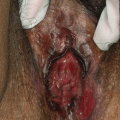
Pyogenic granuloma, otherwise known as lobular capillary hemangioma, is a benign vascular proliferation found on the skin, commonly papular or pedunculated, often with a history of rapid growth within days or weeks (Fig. 12.2). It can be complicated by bleeding, especially if traumatized.
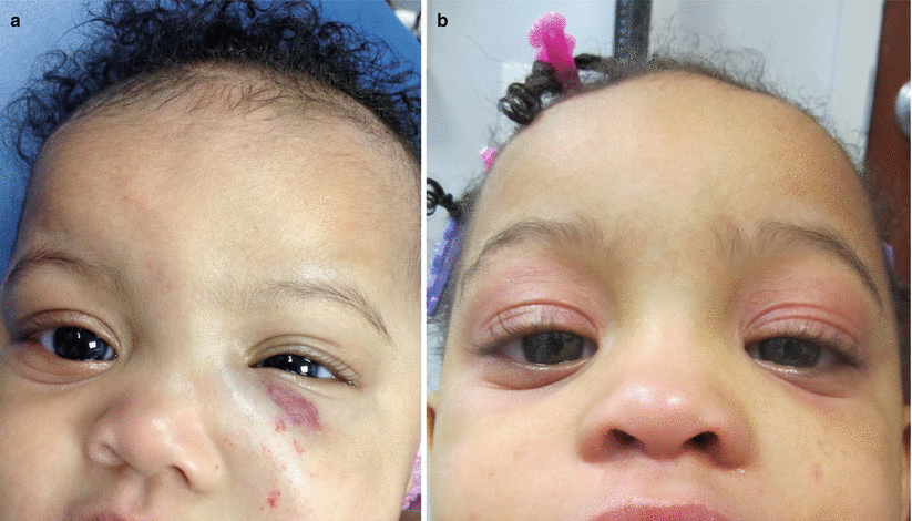
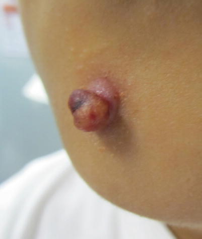
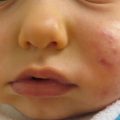
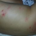
Fig. 12.1
(a) Infantile hemangioma, pre-propranolol; (b) infantile hemangioma, post-propranolol
Fig. 12.2
Pyogenic granuloma
Management Strategies
Treatment of these lesion is mainly surgical. This includes surgical excision, curettage/shave excision +/− cautery, punch biopsy, as well as ligation. Other treatment options include cryotherapy, laser therapy (CO2, pulsed dye laser, Nd-Yag, 1,064 nm), sclerotherapy, and imiquimod 5 % cream [8].
Investigations Recommended
For diagnosis |
Clinical diagnosis or tissue biopsy
Related posts:Stay updated, free articles. Join our Telegram channel
Full access? Get Clinical Tree
 Get Clinical Tree app for offline access

|