Abstract
An infantile hemangioma (IH) represents the most common benign tumor arising in infancy. Adopting specific terminology has enabled IHs to be distinguished from other childhood vascular anomalies, including rapidly involuting and non-involuting congenital hemangiomas (RICH and NICH). The natural history of IHs is characterized by a proliferative phase during the first few months of life, followed by slower involution over a period of years. Variations in the typical growth pattern of IHs can occur, such as lesions with minimal or arrested growth. Cutaneous IHs are characterized as superficial, deep, and mixed lesions based on their clinical appearance. Extracutaneous IHs occasionally occur, such as airway and hepatic hemangiomas associated with cutaneous IHs in a “beard” distribution and multifocal small superficial IHs, respectively. In addition, larger IHs in a segmental pattern can be associated with regional extracutaneous congenital anomalies. Most IHs do not require active treatment and involute without leaving significant residua; however, some IHs are problematic due to complications such as ulceration, interference with a vital function (e.g. vision, patent airway), or potential disfigurement. Early recognition of the IHs at greatest risk of complications is essential for optimal management. Discoveries related to the clinical and immunohistochemical features of IHs and medical therapy with β-blockers have significantly improved the management of complicated IHs.
Keywords
infantile hemangioma, superficial infantile hemangioma, deep infantile hemangioma, segmental infantile hemangioma, rapidly involuting congenital hemangioma (RICH), non-involuting congenital hemangioma (NICH), infantile hemangioma with minimal or arrested growth, PHACE(S), LUMBAR, β-blockers, propranolol, timolol, multifocal infantile hemangiomas, hepatic hemangioma, ulcerated infantile hemangioma
- ▪
The most common benign soft tissue tumor of infancy
- ▪
Demonstrate a typical growth pattern characterized by early proliferation followed by gradual, spontaneous involution
- ▪
Distinct histopathologic and immunohistochemical features that differentiate them from other vascular anomalies in children
- ▪
May be associated with extracutaneous findings when occurring at certain anatomic sites, e.g. the face, neck, lumbosacral area
- ▪
Treatment choice, if any, depends on multiple factors and must be tailored to each individual patient
- ▪
Systemic β-blockers have become the first-line therapy for most children with complicated hemangiomas
Introduction
Infantile hemangiomas (IHs) are benign proliferations of endothelial cells and their supporting tissues and represent the most common tumors arising in the neonatal period. They are characterized by significant growth during the first several months of life, followed by slow spontaneous involution over the ensuing years. This natural history differentiates them from vascular malformations ( Table 103.1 ). Studies highlighting the role of a specific IH stem cell have provided new insights into their pathophysiology.
| DIFFERENCES BETWEEN INFANTILE HEMANGIOMAS AND VASCULAR MALFORMATIONS | ||
|---|---|---|
| Infantile hemangioma | Vascular malformation | |
| Clinical |
|
|
| Epidemiology | More common in:
| No gender or gestation predilection |
| Pathology | Proliferating : endothelial cell hyperplasia, lobule formation, mast cells, prominent basement membrane Involuting : fibrofatty tissue replacement, decreased mast cells when fully involuted | Dependent upon type, often irregular vascular channels |
| Immunohistochemistry | Positive for GLUT1, Lewis Y antigen, merosin, and FcγRII, Wilms tumor protein 1 (WT1) | Negative for GLUT1, Lewis Y antigen, merosin, and FcγRII, WT1 |
History
The terms “infantile hemangioma” and “hemangioma of infancy” describe a specific group of vascular tumors that arise during infancy and demonstrate characteristic clinical and histologic features. IHs have been recognized in the medical literature for centuries, and they have been given various names such as nevus maternus , angioma simplex , angioma cavernosum , angiodysplasia , strawberry nevus , and capillary hemangioma . Despite early attempts to make distinctions among different types of vascular lesions of infancy, the term “hemangioma” was frequently applied in a generic manner to a wide variety of congenital and acquired vascular anomalies including vascular malformations, without consideration of differences in their biologic behavior.
One of the most important developments in the study of vascular “birthmarks”, including lesions that become apparent during early infancy, was the acceptance of a biologic classification scheme. In 1982, Mulliken and Glowacki first proposed that vascular birthmarks should be categorized according to their biologic and clinical behavior. The International Society for the Study of Vascular Anomalies modified this classification system in 1996 and subsequently in 2014 to reflect new insights, including the genetic bases of several types of vascular malformations ( Table 103.2 ) .
| BIOLOGIC CLASSIFICATION OF VASCULAR BIRTHMARKS |
|---|
| Vascular tumors |
Benign
Locally aggressive or borderline
|
| Vascular malformations |
Simple
Combined
Of major named vessels Associated with other anomalies
|
| Provisionally unclassified |
|
Under this organization scheme, vascular birthmarks are divided into two main categories, vascular tumors and vascular malformations, as well as a third group of “provisionally unclassified” lesions for which the biologic behavior has not yet been well characterized. Vascular tumors are characterized by cellular proliferation and include IH, pyogenic granuloma, and less common neoplasms that may arise during infancy or early childhood, including tufted angioma and kaposiform hemangioendothelioma (see Table 103.2 and Ch. 114 ). Vascular malformations, on the other hand, are believed to represent errors in vascular morphogenesis. They are frequently noted during the neonatal period, but, unlike IHs, they do not rapidly proliferate in the first year of life nor do they resolve spontaneously (see Table 103.1 ). Vascular malformations are characterized by the type of dysplastic vessels they contain and by their flow properties (see Ch. 104 ).
Adopting specific terminology has allowed investigators to better categorize vascular birthmarks and predict their clinical behavior and prognosis. The biologic classification was expanded to include histopathologic, immunohistochemical, and genetic features that aid in distinguishing IHs from other vascular lesions. Despite the wide acceptance of the biologic classification scheme, nosologic confusion still persists in the medical community as well as in the literature.
Epidemiology
IHs arise during the first year of life and are the most common tumor of infancy. They develop in 4–5% of infants, with lesions usually noted within the first several weeks of life . IHs may occur more commonly in Caucasian infants than in other racial groups, but this predilection has not been consistently observed . A female : male ratio of 2–5 : 1 has been noted in multiple retrospective studies as well as prospective studies conducted in dermatology practices; however, a recent hospital-based prospective birth-cohort study found no female predominance among infants who developed an IH . A higher female : male ratio of 7–9 : 1 has been reported for patients with severe, complicated IHs.
IHs also develop more frequently in premature infants. Low birth weight represents an independent IH risk factor, and IHs affect 25–30% of infants with a birth weight <1000 g and 15% of those with a birth weight between 1000 and 1500 g . Factors associated with placental insufficiency (e.g. preeclampsia, placenta previa) are also associated with IH development, while prematurity and multiple gestation pregnancies are linked with the development of multifocal IHs . One study found a threefold increased incidence in infants born following chorionic villus sampling compared to those born following amniocentesis or without a history of prenatal instrumentation . A higher maternal age has also been associated with hemangioma development .
IHs typically arise sporadically, although Margileth and Museles reported a 10% incidence of familial cases in their 1965 series. The frequent occurrence of hemangiomas in the general population makes it difficult to assess the true familial incidence, and no specific genes have been consistently implicated.
Pathogenesis
IHs represent localized or regional areas of abnormal vascular development and proliferation. Several hypotheses have been proposed to explain their pathogenesis, but no single theory accounts for all their features. It is likely that several mechanisms under the control of multiple genes, in addition to local effects, play a role in the development, growth, and involution of hemangiomas.
Hemangioma Endothelial Cells: Origin, Signaling Pathways, and Genetics
Hemangioma-derived stem cells (HemSCs) and endothelial progenitor cells have both been isolated from IH specimens. HemSCs can differentiate into endothelial cells, pericytes, and adipocytes. Recent evidence supports derivation of IHs from multipotent stem cells, implying that IH development involves de novo formation of vessels from progenitor/stem cells (vasculogenesis) as well as formation of new vessels from existing ones (angiogenesis) .
The overlapping immunohistochemical phenotype of hemangioma cells and human placental endothelium implies that the former may be of placental origin or undergo differentiation toward a placental microvascular phenotype. North and co-workers reported that glucose transporter protein-1 (GLUT1) is expressed by IHs during all phases of their development (proliferating, involuting, involuted) as well as by the placenta, but not by other vascular tumors or malformations. Additional placenta-associated vascular antigens, including merosin, FcγRII, and Lewis Y antigen, are present in hemangioma specimens and placental chorionic villi but absent in microvessels of the normal skin and subcutis . However, hemangiomas do not express placental trophoblastic markers (e.g. human placental lactogen) and are no longer thought to represent placental emboli.
Several signaling pathways play a role in IH development. Studies have confirmed the importance of vascular endothelial growth factor (VEGF; also known as VEGF-A) signaling, with a shift from expression of VEGF receptor 1 (VEGFR-1), which binds VEGF with high affinity but transmits a very weak signal, to VEGFR-2, which strongly stimulates endothelial cell proliferation upon VEGF binding . VEGFR-2 signaling activates the phosphatidylinositol 3-kinase (PI3K)-mammalian target of rapamycin (mTOR) pathway, leading to upregulation of hypoxia inducible factor-1α (HIF-1α) and increased levels of VEGF. Accordingly, treatment of hemangioma endothelial cells in vitro with the mTOR inhibitor rapamycin results in reduced proliferation . Hemangiomas also have increased expression of the endothelial cell-specific Tie-2 tyrosine kinase receptor and dysregulated production of angiopoietin-2, which may promote angiogenesis and inhibit vessel maturation (see Ch. 102 ) . The Notch pathway is thought to mediate differentiation of HemSCs into pericytes/vascular smooth muscle cells , and the latter appear to have pro-angiogenic properties and support IH development .
A variety of genetic factors may be involved in the development of IH. There have been reports of somatic mutations in genes that encode proteins involved in VEGF signaling (e.g. VEGFRs) and other pathways that affect vascular development within hemangioma tissue . Heterozygous germline mutations in VEGFR2 and ANTXR1 (anthrax toxin receptor 1), which encodes an integrin-like endothelial cell receptor, have been identified in a small subset of IH patients and may represent hemangioma predisposition factors . In addition, familial hemangiomas in several kindreds have been linked to chromosome 5q , and loss of heterozygosity of 5q has been noted in sporadic hemangiomas , suggesting that gene(s) at this locus may be involved in hemangioma formation.
Hypoxia and Other Extrinsic Factors
A role for hypoxia in the pathogenesis of IHs is supported by their association with hypoxic placental changes, prematurity/low birth weight (often caused by placental insufficiency), retinopathy of prematurity (GLUT1-positive, hypoxia-induced neovascularization), and regional arterial insufficiency in PHACE[S] and LUMBAR syndromes (see below) . Hypoxia upregulates expression of GLUT1 and VEGF, leading to mobilization of endothelial progenitor cells. Moreover, the combination of hypoxia and estrogen has a synergistic effect on hemangioma endothelial cell proliferation in vitro , potentially explaining the predilection of IHs for female infants. Investigators have also found that HIF-1α and its downstream effectors are upregulated in IHs during their proliferative phase .
Other cell types in the hemangioma’s environment, including monocytes, fibroblasts, pericytes, mesenchymal cells, adipocytes, and mast cells, can influence endothelial cell proliferation and may have a role in hemangioma formation and/or involution.
Factors Influencing Natural History
Hemangiomas have been studied during different phases of their clinical course to gain insights into the mechanisms that govern their growth and involution. In the proliferative phase, hemangiomas express markers of proliferation such as proliferating cell nuclear antigen (PCNA) and increased levels of pro-angiogenic molecules such as VEGF and basic fibroblast growth factor (bFGF) . In addition, other angiogenesis mediators, proteins found within the perivascular extracellular matrix, and enzymes involved in matrix remodeling are produced at higher levels during proliferation ( Table 103.3 ) .
| MARKERS OF PROLIFERATING AND INVOLUTING HEMANGIOMAS |
|---|
| Proliferative phase |
|
| Involution |
|
* VEGF and bFGF, but not the angiogenesis inhibitor interferon-β, are also expressed by the hyperplastic epidermis overlying proliferating hemangiomas.
The factors that govern the transition of hemangiomas from proliferation to involution and the mechanisms of the associated decrease in pro-angiogenic factors are not fully understood . Increased expression of angiogenesis inhibitors and apoptosis promoters has been observed in involuting hemangiomas (see Table 103.3 ) .
Clinical Features
Presentation
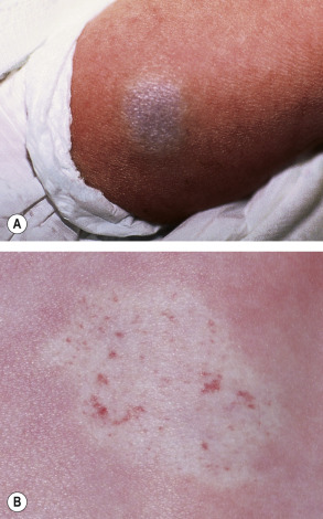
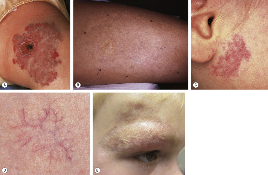
Most IHs have a typical presentation and growth pattern. Lesions usually become apparent during the first few weeks of life, although the proportion of hemangiomas that are “congenital” in published series has ranged from 15% to 60% . IHs evident at birth most often appear as precursor lesions but occasionally they present as relatively well-formed typical IHs that subsequently demonstrate variable proliferation then slow involution; rapidly involuting and non-involuting congenital hemangiomas (RICH/NICH) represent separate entities that are discussed below. Precursor IH lesions include telangiectasias surrounded by a vasoconstricted halo, areas of pallor, pink macules, and blue bruise-like patches ( Fig. 103.1 ). Pink macules and patches may mimic a capillary malformation, and repeat examination over the ensuing weeks is essential to determine the diagnosis ( Fig. 103.2 ). Occasionally, an area of ulceration in the diaper area or on the lip may herald the onset of an IH, and in a newborn infant these lesions may be confused with bacterial or viral infections. Biopsy of the ulcerated lesion may not be clearly diagnostic of an IH, but continued observation usually clarifies the diagnosis .
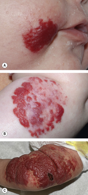
Hemangiomas may occur anywhere on the skin and mucosal surfaces. Although overall they most commonly occur on the trunk , ~50% of the IHs that present to a pediatric dermatologist are located on the head and neck . The clinical appearance of an IH is influenced by its location within the skin and subcutaneous tissues. Superficial hemangiomas are situated in the superficial dermis and are bright red in color during their proliferating phase. The surface is finely lobulated, and the term “strawberry hemangioma” has been used to describe them ( Fig. 103.3 ).
Deep hemangiomas are located in the deep dermis and/or subcutis. They usually are not apparent in the immediate newborn period, often becoming evident a few weeks or even months after birth. They present as warm, ill-defined, light blue–purple masses with minimal or no overlying skin changes, making them more difficult to diagnose than superficial or mixed hemangiomas ( Fig. 103.4 ). The presence of dilated veins or telangiectasias overlying a deep hemangioma provides a clue that the lesion is of vascular origin. Larger deep hemangiomas often have significant arterial blood supply during the proliferative phase, and detection of high flow by Doppler ultrasonography can help to confirm the diagnosis. Mixed hemangiomas have both superficial and deep components, often presenting during the proliferative phase as a well-delineated red vascular plaque overlying a larger, poorly circumscribed violaceous or light blue nodule.
Superficial hemangiomas are the most common type, accounting for ~50–60% of IHs. An additional 25–35% of hemangiomas are mixed and 15% are deep . Approximately 25% of patients have multiple lesions, which are occasionally associated with visceral hemangiomatosis (see below).
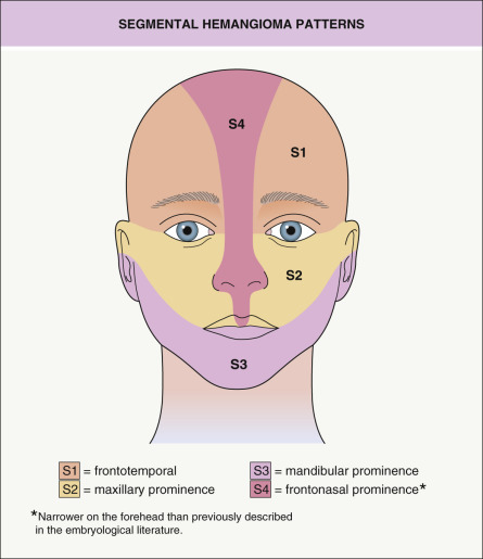
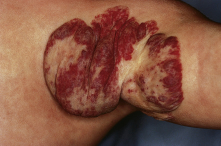
IHs can also be categorized based upon the pattern of involvement, which may help to predict prognosis . The two main pattern subtypes are: (1) focal – arising from a single localized nidus (see Fig. 103.3A, B ); and (2) segmental – covering a broad area or developmental unit in a “plaque-like” manner (see Fig. 103.3C ). Lesions that are difficult to classify are designated as indeterminate . The majority of IHs are focal lesions, and those on the face may show predilection for embryonic fusion lines . The distribution patterns of segmental hemangiomas on the face correlate somewhat with classic embryonic facial prominences, differing the most on the upper face. Investigators have identified four primary segments for facial hemangiomas, S1 to S4 ( Fig. 103.5 ) . Involvement of these segments may be incomplete, and some hemangiomas encompass more than one segment.
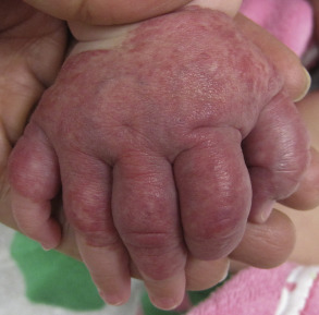
Segmental hemangiomas often begin as broad patches of confluent or reticulated erythema and/or telangiectasias. Within a few weeks, bright red papules and plaques arise within this “field”. Segmental hemangiomas are more likely to be associated with regional extracutaneous anomalies, including PHACE(S) and LUMBAR syndromes (see below) . On the extremities, smaller segmental hemangiomas typically spare the distal digits in a “biker glove” pattern ( Fig. 103.6 ), while larger lesions that encompass the distal digits are associated with a higher risk of extracutaneous anomalies .
Natural History
Studies have documented the typical growth pattern of IHs. The phases include early proliferation with a rapid increase in size, late proliferation with continued growth at a slower rate, plateau (existence as a distinct phase debated), and involution . Hemangiomas tend to “mark out their territory” early on, subsequently undergoing primarily volumetric rather than centrifugal growth . During the proliferative phase, hemangiomas frequently become warmer and firmer in texture, and the surface of superficial hemangiomas may appear tense. Mixed and deep hemangiomas in the proliferative phase often feel firmer and look larger with crying or activity. Deep hemangiomas tend to proliferate for a longer period of time than do superficial hemangiomas, and the deep component of mixed lesions often continues to grow even after the superficial component has plateaued ( Fig. 103.7 ).
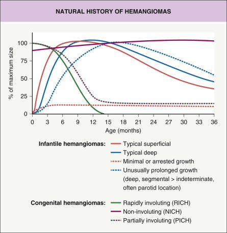
The majority of hemangiomas reach 80% of their final size by the end of the early proliferative phase, which occurs at a mean age of 3 months, and growth is usually most rapid from 5 to 8 weeks of age . The small minority of hemangiomas that grow after 9 months of age tend to have a deep component and/or segmental morphology, and IHs in the parotid gland area are particularly prone to this behavior .
A subset of hemangiomas referred to as infantile hemangioma with minimal or arrested growth (IH-MAG) , “ abortive ”, or “ reticular ” display little or no growth beyond patches of reticulated erythema with coarse and/or fine telangiectasias, often superimposed on a background of pallor ( Fig. 103.8 ) . If present, the proliferative component involves <25% of the surface area, often presenting as small red papules at the periphery . These hemangiomas favor the lower body and may develop recalcitrant ulceration or (if larger and segmental) be associated with LUMBAR or PHACE(S) syndrome (see below) . They are GLUT1-positive and involute at a pace similar to classic IHs, although larger ectatic vessels may persist.
Involution of IHs may begin as early as the first year of life and continues for several years. A color change from bright red to gray–purple ( Fig. 103.9 ) and flattening of the surface are often the earliest signs of involution in superficial lesions. The latter often break apart into smaller islands before clearing. As the tumor involutes, deeper lesions become less firm and the superficial portion may develop a wrinkled appearance. During involution, larger lesions demonstrate less fluctuation of size with crying and activity .
Classic studies on the natural history of untreated hemangiomas demonstrated that 30% of lesions involute fully by 3 years of age, 50% by 5 years, 70% by 7 years, and >90% by 9 years . However, recent studies have concluded that the median age of completed involution is 36 months, with >90% of children showing no further improvement after 4 years of age . Some hemangiomas involute completely, while others leave atrophic, fibrofatty, or telangiectatic residua ( Fig. 103.10 ). Predicting whether the residual lesion will be of cosmetic significance is one of the most challenging aspects of hemangioma management.
Complications
The majority of hemangiomas are small lesions that require little or no intervention. However, some hemangiomas are problematic due to their size, location, or association with other anomalies. The age of the patient and the growth pattern of the hemangioma are crucial factors in predicting complications.
Ulceration
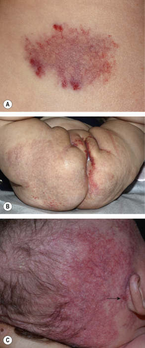
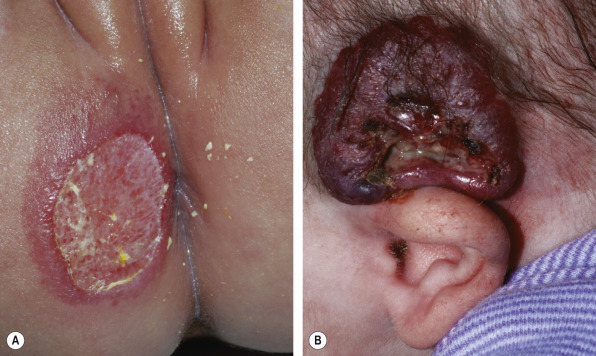
Ulceration occurs in up to 10% of all IHs and represents the most common complication . Although hemangiomas in any location can be affected, those on the lip and in the anogenital region or other skin folds (e.g. the neck) have the greatest tendency to ulcerate ( Fig. 103.11 ). Ulceration develops more commonly within large, mixed (superficial and deep), or segmental hemangiomas . For example, ulceration occurs in up to 30% of segmental extremity lesions (see Fig. 103.3C ) . In addition, IHs-MAG are prone to ulceration, which may be recalcitrant.
Overall the median age at ulceration is 4 months , although whitish discoloration of hemangiomas in infants younger than 3 months of age may be a sign of impending ulceration . In addition to causing pain, ulcers increase the risk of infection and result in scarring with textural change. Bleeding rarely occurs and can usually be controlled with firm pressure. The management of ulcerated hemangiomas may be challenging (see below) .
Disfigurement and interference with function
Large hemangiomas can distort tissues, interfere with function, and lead to significant long-term sequelae. In addition, regional extracutaneous abnormalities may occur, primarily in association with segmental hemangiomas (see below). High-output congestive heart failure represents a rare complication that is more frequently observed in patients with visceral involvement.
Disfigurement and interference with function because of location
Even small lesions may cause complications if they arise in vulnerable locations. Periocular hemangiomas are often associated with ophthalmologic complications ( Fig. 103.12 ). Most commonly, periocular hemangiomas cause astigmatism by compressing the globe and deforming the cornea, which results in asymmetric refractive errors. They may also cause visual abnormalities by obstructing the visual axis or by invading the orbital musculature, which can lead to light-deprivation amblyopia and strabismus, respectively. Lesions located on the upper lid are most problematic, but visual obstruction can complicate lesions on the lower lid as well. Proptosis is a rare presentation of an orbital hemangioma and may develop gradually or suddenly, potentially leading to corneal exposure; the diagnosis of hemangioma may be difficult clinically when a cutaneous lesion is not present. Infants with periocular hemangiomas should be evaluated by an ophthalmologist at baseline and closely thereafter during the proliferative stage .
Hemangiomas on the nasal tip are particularly challenging. Deep and mixed hemangiomas can distort the underlying cartilage and leave significant fibrofatty residua. The resultant “Cyrano-nose” deformity may require reconstructive surgery. In rare cases, superficial hemangiomas located along the columella may ulcerate and lead to destruction of underlying cartilage, which may be heralded by the appearance of a horizontal crease in the inferior columella.
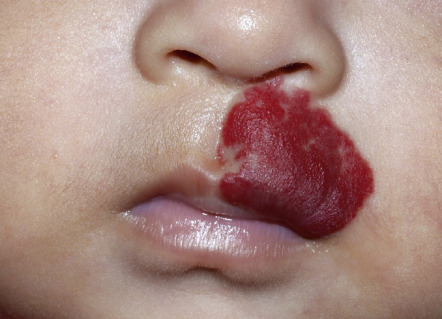
Lip hemangiomas are often superficial or mixed lesions; painful ulceration is common as they proliferate, which leads to feeding difficulties. Local factors, including recurrent trauma and the commensal bacterial flora, may increase the incidence of this complication. Distortion of the vermilion border or lengthening of the lip can result in significant cosmetic residua, eventually requiring surgical correction ( Fig. 103.13 ) .
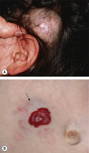
Hemangiomas located on the pinna may ulcerate and become infected, increasing the risk of scarring and distortion of normal structures. Conductive hearing loss can result from obstruction of the external auditory canal by a hemangioma .
Hemangiomas located on the breast pose a particular challenge in girls. These lesions may affect the underlying breast bud, and residual masses may lead to the appearance of breast asymmetry. Early surgical intervention is not advised, as it may ultimately affect normal breast development.
Anogenital hemangiomas are often complicated by painful ulceration, and wound care may be difficult in this region. Large hemangiomas of the limbs may also ulcerate, and residual excess tissue and rarely limb-length discrepancy can remain upon involution.
Extracutaneous involvement
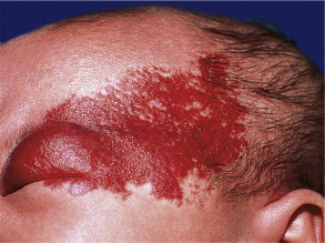
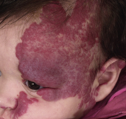
Large facial hemangiomas, specifically segmental lesions >5 cm in diameter, can pose challenges beyond the complications noted above. They are often associated with extracutaneous anomalies, especially in female infants . These hemangiomas may present as segmental telangiectatic patches (e.g. IH-MAG), vascular plaques, or deep masses. A relationship between facial hemangiomas and structural or vascular CNS anomalies was recognized >35 years ago . Since then, other associated congenital anomalies have been identified. In 1996 , the acronym PHACE(S) syndrome was coined for this spectrum of findings: P, p osterior fossa and other structural brain malformations; H, h emangioma; A, a rterial anomalies of cervical and cerebral vessels; C, c ardiac defects (especially c oarctation of the aorta); E, e ye anomalies; and S, s ternal defects and s upraumbilical raphe ( Fig. 103.14 ). Occasionally, impaired hearing and endocrine abnormalities such as hypopituitarism and hypothyroidism are additional features . Diagnostic criteria for PHACE(S) syndrome were established by a multidisciplinary group of specialists in 2009 and updated in 2016 ( Table 103.4 ).
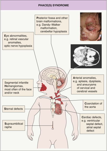
| DIAGNOSTIC CRITERIA FOR PHACE(S) SYNDROME | ||
|---|---|---|
Definite PHACE(S):
Possible PHACE(S):
| ||
| Organ system | Major criteria | Minor criteria |
| Arterial |
| |
| Structural brain |
|
|
| Cardiovascular |
|
|
| Ocular |
|
|
| Ventral or midline |
|
|
* Includes internal carotid artery; middle, anterior, or posterior cerebral artery; and vertebrobasilar system.
** Includes kinking, looping, tortuosity, and/or dolichoectasia.
In a prospective study of 108 infants (age <1 year; mean age 3 months) with large facial hemangiomas (≥22 cm 2 ), 31% had PHACE(S) syndrome. The vast majority (91%) of affected individuals had more than one extracutaneous manifestation (major ± minor criteria), and the most common findings were cerebrovascular (91%), cardiovascular (67%), and structural brain (52%) anomalies . The cerebrovascular anomalies typically occur ipsilateral to the hemangioma and most commonly involve the internal carotid artery, highlighting the need for both head and neck imaging . Aberrant subclavian artery and coarctation of the aorta, each present in ~20% of infants with PHACE(S), represent the most frequent cardiovascular anomalies .
An approach to the evaluation of infants at risk for PHACE(S) syndrome is presented in Fig. 103.15 , and consensus-derived recommendations for evaluation and ongoing care were recently published . Cerebrovascular and cardiovascular changes may be progressive, with potential complications including ischemic stroke , and affected infants should be followed by neurologists and cardiologists as clinically appropriate.
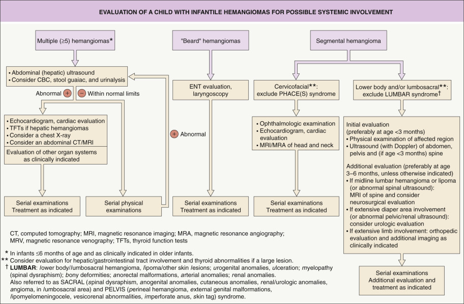
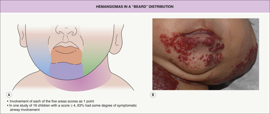
Lower facial or “beard” hemangiomas often serve as markers of laryngeal hemangiomatosis, and the risk can be estimated by the extent of cutaneous involvement in this region ( Fig. 103.16 ) . However, airway hemangiomas are occasionally associated with segmental hemangiomas that primarily involve the upper face or small hemangiomas in the “beard” area . Airway hemangiomas are typically subglottic. The onset of symptoms, which include noisy breathing and biphasic stridor, ranges from a few weeks to several months of age . Infants with lower facial hemangiomas of concern should be referred for otolaryngologic evaluation.
Related posts:


Stay updated, free articles. Join our Telegram channel

Full access? Get Clinical Tree


