Abstract
The evaluation of a patient with hair loss requires a detailed history, physical examination, and, in some cases, laboratory tests and biopsy ( Table 20.1 ). Important elements of the history include the time of onset, medications taken, recent emotional or physical stress, diet, grooming techniques, and family history of baldness or hair disorders.
The physical examination is helpful in making an accurate diagnosis by observing the pattern (patchy or diffuse) of hair loss and whether scarring is present as evidenced by loss of follicular openings. Patchy hair loss is readily apparent. However, diffuse hair loss may not be noticeable until the patient has more than 50% hair loss. The presence or absence of scarring is important diagnostically and prognostically. In nonscarring alopecia, the diagnosis is usually made without biopsy. In scarring alopecia, a biopsy is useful in establishing a prognosis and diagnosis and should be performed. Nonscarring alopecia may be a temporary phenomenon, whereas scarring indicates permanent hair loss. Except for discoid lupus, the main disorders discussed in this chapter are nonscarring.
Alopecia Areata
- 1.
Autoimmune disorder
- 2.
Acute onset of well-circumscribed, oval patches of nonscarring alopecia
- 3.
No cure
| Incidence (%) a | History | Physical Examination | Scarring | Pattern | Differential Diagnosis | Laboratory Test (Biopsy) | |
|---|---|---|---|---|---|---|---|
| Alopecia areata | 0.9 | Acute onset | Exclamation point hairs | Absent | Circular patches | Trichotillomania Secondary syphilis Fungal infection | None |
| Discoid lupus erythematosus | < 0.1 | Photosensitivity Other symptoms of lupus | Erythema Follicular plugs | Present | Patchy | Fungal infection Lichen planopilaris Neoplasm | Biopsy |
| Androgenetic (Male & female pattern hair loss) | 0.6 | Family history of thinning | Normal scalp | Absent | Patterned | Androgen excess in women | None |
| Telogen effluvium | 1.0 | Physical or emotional stress 2–3 months previously | Positive hair pull > 25% telogen hair | Absent | Diffuse | Female pattern hair loss Identify trigger or cause | None |
| Trichotillomania | 0.1 | Emotional problems | Broken hair | Absent | Patchy | Alopecia areata Tinea capitis | None |
| Tinea capitis | 0.1 | Schoolmates with hair loss | Scaling, erythema, pustules | Absent | Patchy | Seborrheic dermatitis Alopecia areata Bacterial infection Trichotillomania | KOH preparation, culture |
a Percentage of new dermatology patients with this diagnosis seen at the Hershey Medical Center Dermatology Clinic, Hershey, PA.
Definition
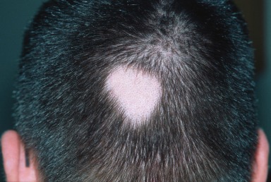
Alopecia areata is an idiopathic disorder characterized by well-circumscribed, round or oval patches of nonscarring hair loss ( Fig. 20.1 ).
Incidence
Alopecia areata affects both sexes equally, with onset occurring most often in early adulthood. Almost 1% of the authors’ new patients had this diagnosis. The incidence in Olmsted County, Minnesota, was 20.2 per 100,000 person-years. Alopecia areata occurs in 1.7% of Americans by the age of 50 years.
History
Alopecia areata has an acute onset. It is sometimes associated with emotional stress, but in most patients the emotional stress seems to be caused by the hair loss. Approximately 25% of patients have other autoimmune disorders, such as type 1 diabetes mellitus, thyroid disease, and vitiligo. Atopic dermatitis is especially common in alopecia areata. Patients are generally healthy otherwise. Some 20% to 25% of patients have a family history of alopecia areata.
Physical Examination
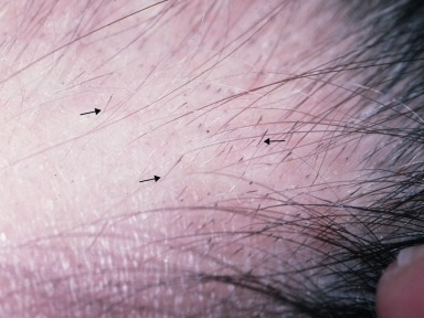
The disorder is characterized by well-circumscribed, round or oval patches of hair loss, leaving a smooth, normal- appearing scalp. Erythema and slight tenderness may be present early in the course. Characteristically, the periphery of the patches of hair loss is studded with exclamation point hairs , which are so named because of their resemblance to a punctuation mark ( Fig. 20.2 ). These fractured hairs are 2 to 3 mm long and tapered at the base.
Alopecia areata is characterized by nonscarring circular patches of alopecia with exclamation point hairs.
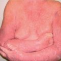
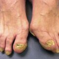
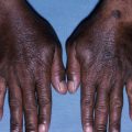
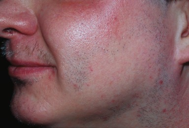
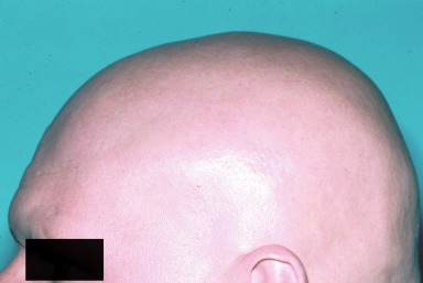
Alopecia areata most often affects the scalp, frequently with several 2- to 3-cm patches of hair loss. The eyebrows, eyelashes, and beard may also be affected, as may hair elsewhere on the body ( Fig. 20.3 ). Approximately 1% to 2% of patients develop loss of all scalp hair ( alopecia totalis ) or loss of all body hair ( alopecia universalis ) ( Fig. 20.4 ). Fine stippling and pitting of the nails are infrequent associated findings.
Differential Diagnosis
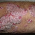
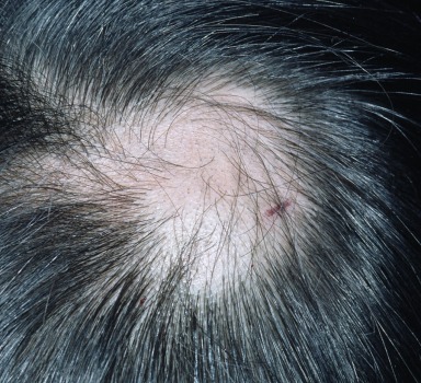
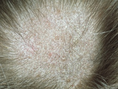
Other nonscarring forms of alopecia need to be considered in the differential diagnosis. Secondary syphilis can be ruled out by appropriate serologic examination. Trichotillomania and tinea capitis should also be considered. Ill-marginated, irregular patches of alopecia containing the stubble of broken hairs are typical of trichotillomania ( Fig. 20.5 ). If doubt exists, a biopsy helps to differentiate trichotillomania from alopecia areata. A potassium hydroxide (KOH) preparation and culture, and clinical evidence of redness and scale, enable the diagnosis of a fungal infection ( Fig. 20.6 ).
- ●
Secondary syphilis
- ●
Trichotillomania
- ●
Tinea capitis
Laboratory and Biopsy
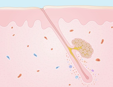
Histopathologic examination of alopecia areata reveals the presence of small, dystrophic hair structures. A lymphocytic infiltrate surrounds the early anagen hair bulbs like a “swarm of bees” ( Fig. 20.7 ).
Therapy
The treatment of alopecia areata depends on the extent of involvement and the patienťs emotional need for regrowth of hair. There is no cure for alopecia areata. In localized disease, topical potent steroids such as clobetasol (Temovate) gel or intralesional injections of triamcinolone (Kenalog-10) are sometimes effective. In widespread disease, systemic steroids are sometimes used, but their hazards must be considered before starting treatment. Prompt hair loss after discontinuation of oral steroids is discouraging. Other modes of therapy include immunotherapy by induction of allergic contact dermatitis (aka contact sensitization), phototherapy, topical minoxidil, and oral cyclosporine. Janus kinase (JAK) inhibitors, both orally and topically administered, are under investigation. Patients with alopecia areata need psychologic support, and all patients should visit the National Alopecia Areata Foundation’s website ( http://www.naaf.org ) or Children’s Alopecia Project ( www.childrensalopeciaproject.org ). A wig is recommended when the hair loss is extensive.
Course and Complications
Alopecia areata has a variable, unpredictable course. Most patients with localized disease have spontaneous recovery. However, relapses are not uncommon. Duration of more than 1 year and extensive hair loss are poor prognostic signs. Spontaneous regrowth of hair in alopecia totalis (scalp) and alopecia universalis (total body) may occur but is uncommon; fewer than 5% of patients show any tendency toward hair regrowth.
Poor prognosis:
- 1.
Long duration
- 2.
Large areas of alopecia
Pathogenesis
The pathogenesis of alopecia areata remains poorly understood, although an immunologic process is favored. Recent research shows the common initiation of the autoimmune response in alopecia areata, celiac disease, rheumatoid arthritis, and diabetes. A lymphocytic inflammatory infiltrate surrounds the affected bulbs and presumably has a role in the disease. In response to this autoimmune process, the hair matrices become arrested, but retain the capacity for normal hair regrowth after months or years.
Lupus Erythematosus
- 1.
Alopecia can be scarring or nonscarring
- 2.
Biopsy confirms diagnosis
- 3.
Treat aggressively to prevent permanent hair loss
Definition
Lupus erythematosus is an autoimmune disorder that often affects the scalp and causes alopecia. Hair loss may be diffuse and nonscarring (systemic lupus erythematosus; SLE) or patchy and scarring (discoid lupus erythematosus; DLE). A general discussion of lupus erythematosus is found in Chapters 9 and 14 .
Physical Examination
Diffuse nonscarring alopecia of the scalp in the form of a telogen effluvium accompanies the acute phases of SLE in more than 20% of patients. In addition, short, broken hairs (“lupus hair”) may be present, particularly in the frontal margin.
SLE – nonscarring
DLE – scarring
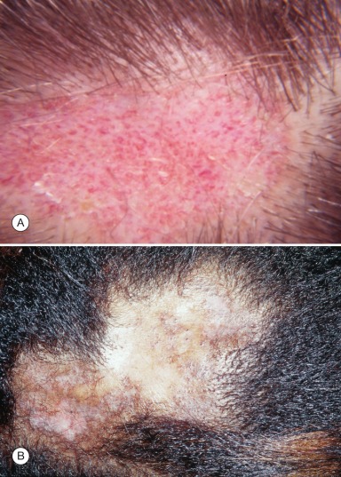
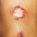
Discoid lupus erythematosus is characterized by oval, scarring areas of alopecia. A typical plaque has an active erythematous margin and a white, atrophic, inactive center ( Fig. 20.8 ). Within the plaques, telangiectasia and dilated keratin-filled follicles are present. Similar discoid lesions may be found on the ears, face, trunk, and extremities.