Abstract
When implanted or injected into the skin, inorganic and high-molecular-weight organic materials can elicit a range of inflammatory reactions, from granulomatous to lichenoid to pseudolymphomatous. Less commonly, reactions resemble photoallergic dermatitis or pyogenic granulomas. These “foreign bodies” can be introduced accidentally (e.g. via motor vehicle accident) or purposefully (e.g. cosmetic tattoos, soft tissue fillers). Occasionally, agents applied topically such as zirconium oxide or paraffin may lead to a foreign body reaction.
Most commonly, patients develop granulomatous foreign body reactions characterized clinically by red to red–brown papules, nodules and/or plaques which may ulcerate. Histologically, there are several types of granulomatous reaction patterns – foreign body, sarcoidal, and palisading. Less often, the histologic pattern is lichenoid, eczematous or pseudolymphomatous. Treatment options are intralesional corticosteroids and surgical excision, if possible.
Keywords
foreign body reaction, granulomatous foreign body reaction, lichenoid foreign body reaction, pseudolymphomatous foreign body reaction, pseudolymphomas, foreign body granulomas, tattoo, paraffin, silicone, silica, soft tissue fillers
- ▪
Foreign body reactions represent inflammatory responses to inorganic and high-molecular-weight organic materials that have been introduced into the skin and are variably resistant to degradation
- ▪
Routes of introduction can vary from accidental or self-inflicted to surgical and cosmetic procedures to topical application of various compounds
- ▪
The most common clinical presentation is red to red–brown papules, nodules or plaques (with or without ulceration) that are due to granulomatous inflammation
- ▪
Less common clinical presentations include lichenoid and pseudolymphomatous lesions, or the development of a fistula or draining wound
- ▪
Histopathologic findings vary from granulomas (foreign body, sarcoidal, palisading) to lichenoid, eczematous or pseudolymphomatous patterns
- ▪
Diagnosis of foreign body reactions requires a high index of suspicion, thorough history, recognition of distinctive distribution patterns, and histologic examination (including polarized microscopy)
General Considerations
Definition
Literally, any material – living or non-living – introduced into the body is a “foreign body” and is treated by our immune system as “non-self” in order to elicit an appropriate response. Such a broad definition also includes infectious agents, but they are covered elsewhere (see Chapter 74 , Chapter 75 , Chapter 76 , Chapter 77 , Chapter 78 , Chapter 79 , Chapter 80 , Chapter 81 , Chapter 82 , Chapter 83 , Chapter 84 , Chapter 85 ). This chapter will focus on non-living materials that have been introduced into the dermis or subcutis . They represent inorganic compounds or organic material of high molecular weight (or their products) that resist degradation by the body’s inflammatory cells. Although granulomatous reaction patterns are most commonly observed, there is a broad range of clinicopathologic presentations. Foreign bodies reported to elicit skin reactions are listed in Table 94.1 .
| CLASSIFICATION OF FOREIGN BODIES ACCORDING TO THEIR ORIGINS AND ROUTES OF ENTRY | ||
|---|---|---|
| Origin | Route of entry | |
| Non-biologic (inorganic/metallic compounds) | ||
| Tattoo inks | Decoration Cosmetic Accidental Iatrogenic | |
| Ferrous subsulfate (Monsel’s solution) | Hemostasis (iatrogenic) | |
| Paraffin | Tissue augmentation Iatrogenic | |
| Silicone (polydimethyl siloxane) liquid or gel | Tissue augmentation | |
| Silica | Wound contamination Blast injuries | |
| Talc | Surgical procedures Wound contamination Umbilical stump contamination Intravenous injection of self-prepared drugs | |
| Zirconium | Topical application: antiperspirants, antipruritic medications | |
| Beryllium | Laceration by broken fluorescent lamps (historical) Penetration of intact skin by fine particles (<1 micron) Inhalation | |
| Aluminum | Subcutaneous injection of aluminum-containing vaccines | |
| Zinc | Subcutaneous injection of insulin–zinc preparations | |
| Other synthetic fillers, e.g. poly-L-lactic acid, calcium hydroxylapatite, polymethylmethacrylate | Tissue augmentation | |
| Biologic | ||
| Produced in the skin but normally isolated from body’s immune surveillance | Hair keratin (see Ch. 38 ) | Ruptured follicle or cyst * Ingrown hair Interdigital pilonidal sinus (barber’s hair sinus) |
| Nail keratin | Ingrown nail | |
| Produced by other living organisms | Starch | Surgical procedures |
| Cactus spines | Accidental Occupational | |
| Jellyfish and corals | Accidental Swimming or diving | |
| Sea urchin spines (see Ch. 85 ) | Accidental Swimming or diving | |
| Silk sutures | Surgical procedures | |
| Hyaluronic acid | Tissue augmentation | |
| Bovine collagen | Tissue augmentation | |
| Miscellaneous | Corticosteroids | Intralesional injection |
| Sutures, primarily absorbable (rarely nylon or polypropylene) | Surgical procedures | |
| Polyamide synthetic “hair” fibers | Scalp “hair” restoration | |
| 2-octyl cyanoacrylate | Tissue adhesive for small, superficial wounds and lacerations | |
| Hydrophilic polymer and potassium ferrate (WoundSeal ® powder) | Hemostasis | |
Routes of Entry
Accidental
Inoculation with foreign bodies may occur unintentionally during various activities such as gardening (wood splinters, cactus spines) and swimming and diving (coelenterate envenomation, sea urchin spines), or it may result accidentally, such as in a blast injury (silica particles) or motor vehicle accident.
Surgical procedures
Contamination of wounds by talc or starch powders used for lubrication of surgical gloves may incite a foreign body reaction. Surgical sutures can also cause a foreign body response (see Fig. 151.19 ). Histologically, suture granulomas are commonly seen in excisional specimens of previously sampled cutaneous tumors. More recently, foreign body reactions have been observed in association with 2-octyl cyanoacrylate, a tissue adhesive used for small superficial wounds and lacerations, as well as a copolymer-containing lubricating agent (Carbopol 934) used for liposuction .
Iatrogenic
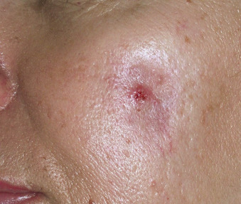
Foreign material implanted into the skin for the purpose of tissue augmentation, e.g. injectable soft tissue fillers, can induce reactions in some individuals ( Fig. 94.1 ). Paraffin is still being used illegally for tissue augmentation, and a paraffin foreign body reaction can also occur after the application of paraffin-containing materials such as nasal packs .
Tattooing
Tattooing is the introduction of an exogenous pigment into the dermis, either deliberately or accidentally, resulting in permanent discoloration of the skin.
Topical application
Antiperspirant and antipruritic preparations containing zirconium oxide applied to the skin surface may cause foreign body reactions. Of note, zirconium lactate was generally banned in 1978.
Self-inflicted
Self-administered intravenous injections, a common practice among drug addicts, may lead to the introduction of foreign material into the skin.
Pathogenesis
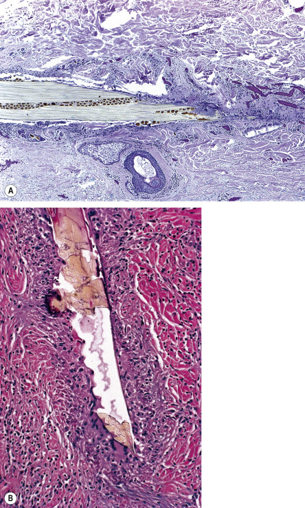
The initial tissue response to most foreign substances involves an accumulation of neutrophils, which usually fail to properly deal with the foreign body. Its persistence attracts monocytes and local tissue macrophages that engulf the foreign material and become activated. Engulfed material may resist degradation and remain sequestered within the macrophages. Activated macrophages secrete a variety of specific biologically active substances, e.g. cytokines that attract additional macrophages and blood monocytes. The formation of a chronic granuloma represents an attempt by the body to sequester a persistent indigestible material. Individual macrophages may become larger (epithelioid histiocytes) or fuse to form multinucleated foreign body giant cells ( Fig. 94.2 ). The infiltrate also contains T lymphocytes and fibroblasts. The pathogenesis of other patterns of reactions, e.g. lichenoid, pyogenic granuloma-like, pseudolymphomatous, remains speculative.
Clinical Features
The host response and, consequently, the clinical presentations of foreign body implantation are variable ( Tables 94.2 & 94.3 ). An acute inflammatory response occurs shortly after the entry of the foreign material. This may resolve, to be followed weeks, months or even years later by a chronic inflammatory response. Although the latter may have a variety of clinical presentations, red to red–brown papules, nodules and plaques (with or without ulceration) are most commonly observed. Over time, the lesions may become firmer due to fibrosis. The pattern and arrangement of the cutaneous lesions will correspond to the route of inoculation, and observation of this patterning, in association with a relevant patient history, is crucial.
| CLINICAL PRESENTATIONS OF FOREIGN BODY REACTIONS | |
|---|---|
| Clinical presentation | Foreign substance |
| Erythema, induration, papules, nodules, plaques (with or without ulceration) | Tattoo inks, paraffin, silicone * , silica * , beryllium (local), starch, soft tissue fillers (e.g. hyaluronic acid) |
| Sarcoidosis-like granulomatous papules | Tattoo inks, talc, zirconium, beryllium (systemic), soft tissue fillers (e.g. hyaluronic acid, bovine collagen) |
| Pyogenic granuloma-like lesions | Talc, keratin (ingrown nail) |
| Pseudofolliculitis barbae, “ingrown” hairs of the pubic region and upper thighs | Keratin |
| Pruritic lichenoid papules and plaques (often in an irregular linear pattern) | Tattoo inks, jellyfish and coral stings |
| Abscess | Keratin (e.g. pilonidal sinus), zinc, paraffin, soft tissue fillers |
| Sinus tract/fistula | Suture, keratin (pilonidal sinus, barber’s sinus when interdigital) |
| Persistent subcutaneous nodules at injection site | Aluminum, soft tissue fillers (see Table 94.4 ) |
| Pseudolymphomatous reaction | Aluminum, tattoo inks |
| Photoallergic eczematous reaction | Tattoo ink containing cadmium sulfide (yellow), cadmium selenide (red), or azo dyes (yellow, red) |
| CLINICAL AND HISTOPATHOLOGIC FEATURES OF FOREIGN BODY REACTIONS | ||||
|---|---|---|---|---|
| Foreign body | Clinical presentations | Histologic reaction patterns | Other distinctive features | Birefringent |
| Tattoo inks | Erythema, induration, papules, nodules Lichenoid papules and plaques Eczematous dermatitis (including photoallergic reactions) | Foreign body or (with certain dyes) sarcoidal granulomas Lichenoid dermatitis Spongiotic dermatitis Pseudolymphoma | Pigment granules scattered throughout the infiltrate, extra- and intracellular (macrophages) | |
| Silica | Nodules, indurated plaques within scar Disseminated papules (blast injuries) Prolonged incubation period | Granulomas (sarcoidal > foreign body) | Colorless crystals, extra- and intracellular | √ |
| Talc | Sarcoid-like papules Thickening and erythema of an old scar Involvement of intertriginous zones, iv injection sites Umbilical stumps Pyogenic granuloma-like | Sarcoidal or foreign body granulomas Pyogenic granuloma-like | Needle-shaped or round talc crystals: clear, blue–green or yellow–brown | √ (“Maltese cross”-like appearance) |
| Zirconium | Persistent, soft, brownish papules Involvement of axillary skin | Sarcoidal granulomas | ||
| Beryllium, local skin reaction | Nodule, ulcer | Caseating granulomas | ||
| Beryllium, systemic | Widely scattered papules (<1% of cases) | Sarcoidal granulomas | ||
| Aluminum | Persistent subcutaneous nodules at injection site | Granulomas with peripheral lymphocytes and eosinophils Pseudolymphoma | Basophilic granular material within the cytoplasm of histiocytes | |
| Zinc | Furuncles at injection sites | Early: dense neutrophilic infiltrate Late: granulomas and fibrosis | Rhomboidal crystals | √ |
| Starch | Papules, nodules | Foreign body granulomas | Ovoid basophilic starch granules PAS-positive | √ |
| Cactus spines | Dome-shaped, skin-colored papules with a central black dot | Early: neutrophils Late: sarcoidal or foreign body granulomas | Spines, extra- and intracellular (giant cells) PAS-positive | √ |
| Jellyfish, corals, sea urchin spines | Pruritic lichenoid papules and plaques (onset 2–3 weeks after exposure) Linear, zig-zag and whip-like (flagellate) patterns of erythema/edema (early), hyperpigmentation or lichenoid papules (late) | Lichenoid dermatitis | Calcite crystals (sea urchin spines) | √ (sea urchin spines) |
| Keratin | Pseudofolliculitis/acne keloidalis Pyogenic granuloma-like lesions, ingrown nails Pilonidal sinus | Foreign body granulomas | Other inflammatory cells | √ |
| Intralesional corticosteroids | Skin-colored to yellow–white papules or nodules develop at the site of a previous intralesional injection of corticosteroid The incubation period varies between weeks to months | Foreign body granulomas | Pale bluish material in H&E-stained sections | |
| Suture | Wound appears inflamed, red, edematous (or develops papules or nodules) and opens to form a fistula | Foreign body granulomas | √ | |
In addition to nodules and plaques, other forms of foreign body reaction include pyogenic granuloma-like lesions , lichenoid lesions , and a chronic draining fistula or wound .
Pathology
Apart from the acute reaction to the trauma that accompanies the introduction of the foreign body, a chronic reaction is the usual response. Although different patterns of chronic local tissue reactions have been described, including lichenoid , eczematous and pseudolymphomatous , the most common is the granulomatous type of reaction (see Table 94.3 ). Granulomas that develop as a reaction to foreign bodies are of two major types:
- •
allergic (immunologic)
- •
non-allergic (“foreign body”).
A histologic decision as to whether a granuloma is of the foreign body type or of the allergic type is not always possible and different patterns may be seen in the same section. It is noteworthy that foreign body reactions in HIV-infected patients are different, with abundant individual macrophages, but no giant cells, suggesting an immunologic dysfunction . The foreign material inciting a reaction may be detected in conventional H&E-stained sections or may require special procedures for its identification (see Table 94.3 ).
Diagnosis
Foreign body reactions should be considered in the differential diagnosis of localized inflammatory nodules and plaques, especially when there is a persistent draining wound or sinus. Occasionally, these reactions present as pyogenic granulomas or localized lichenoid papules. Clinical morphology is usually not distinctive, but there may be a suggestive distribution pattern, and a thorough history is indispensable for arriving at the correct diagnosis.
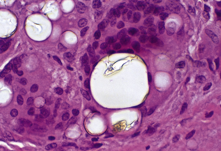
Histopathologic examination can confirm the granulomatous nature of a lesion and occasionally foreign bodies may show distinctive microscopic features in H&E-stained sections ( Fig. 94.3 ) . However, special procedures are usually required for an accurate diagnosis, particularly microscopic examination with polarized light ( Fig. 94.4 ). In general, imaging techniques such as plain radiography, ultrasonography, CT scanning and MRI are of little practical value in visualizing small cutaneous foreign bodies, even if the foreign body is radio-opaque. However, high frequency ultrasound (20 MHz) may be helpful in detecting inflammatory reactions to tattoos and soft tissue fillers, with ultrasound findings correlating with the histopathology of tattoo reactions .