Anesthesia in Patients With Traumatic Facial Injury ( Chapter 1.1 )
Clinical Question:
What are the comparative benefits and harms of general and local anesthesia in patients with traumatic facial injury?
Author Recommendation:
In adults with acute facial trauma, in order to improve patient satisfaction and reduce pain and adverse effects, anesthesiologists, surgeons, and patients should participate in shared decision-making regarding selection of local or general anesthesia based on fracture localization, severity, comorbidities, and individual patient preferences.
What Does the Evidence Conclude?
| Quality of Evidence a | Strength of Recommendations b | Evidence |
|---|---|---|
| Low | Weak | Nasal bone fractures Evidence suggests that general anesthesia is associated with greater patient satisfaction with anesthesia and patient satisfaction with appearance of the nose when compared with local anesthesia in adults with acute nasal bone fractures. The evidence regarding outcomes in patients with different demographics, fracture severity, and comorbidities as well as provider characteristics is insufficient. |
| Very low | Weak | Mandibular fractures Evidence suggests that there are no differences in patient or surgeon satisfaction between general and regional anesthesia in adults with acute mandibular fractures. However, regional anesthesia reduces the duration of pain and need for rescue analgesics and decreases the risk of postoperative nausea, vomiting, and sore throat when compared with general anesthesia. The evidence regarding outcomes in patients with different demographics, fracture severity, and comorbidities as well as provider characteristics is insufficient. |
a Quality of Evidence scale (GRADE): high, moderate, low, and very low. For more information on the GRADE rating system, see http://gdt.guidelinedevelopment.org/app/handbook/handbook.html
b The Guideline Elements Model: http://gem.med.yale.edu.easyaccess1.lib.cuhk.edu.hk/default.htm .
What Are the Parameters of Our Evidence Search?
| PICO | |
|---|---|
| Population |
|
| Intervention |
|
| Comparator |
|
| Primary outcome(s) |
|
Basis for and Determinants of the Strength of Recommendations:
Population:
Adults with acute nasal bone fractures
Setting:
Inpatient
Intervention:
General anesthesia
Comparator:
| Outcomes | Risk With Intervention Per 1000 | Risk With Comparator Per 1000 | Relative Measure of Association (95% Confidence Interval) | Number of Participants (Studies) | Quality (GRADE) | Comments |
|---|---|---|---|---|---|---|
| Patient satisfaction with anesthesia | 838 | 917 | RR 0.9 (0.8;1.0) | 140 (1 RCT) | Very low | No difference |
| Patient satisfaction with anesthesia | 386 | 379 | RR 1.2 (1.0;1.3) | 427 (5 observational studies) | Low | Favors general anesthesia |
| Patient satisfaction with function of the nose | 38 | 25 | RR 1.5 (0.4;6.2) | 248 (2 observational studies) | Low | No difference |
| Patient satisfaction with appearance of the nose | 141 | 44 Attributable events per 1000 treated 98 (2;194) | RR 3.3 (1.4;7.9) NNT 10 (5;500) | 277 (3 observational studies) | Low | Favors general anesthesia |
| Patient preference for treatment if the nose were to refracture | 789 | 879 | RR 0.8 (0.6;1.2) | 189 (2 RCTs) | Very low | No difference |
| Patient preference for treatment if the nose were to refracture | 331 | 305 | RR 1.0 (0.4;2.5) | 248 (2 observational studies) | Very low | No difference |
| Subsequent corrective surgeries Subgroup: septoplasty | 45 | 85 | RR 0.5 (0.2;1.1) | 437 (4 observational studies) | Low | No difference |
| Subsequent corrective surgeries Subgroup: septorhinoplasty | 323 | 257 | RR 1.3 (0.7;2.1) | 139 (1 RCT) | Very low | No difference |
| Subsequent corrective surgeries Subgroup: septorhinoplasty | 33 | 67 | RR 0.6 (0.1;3.3) | 466 (5 observational studies) | Low | No difference |
| Subsequent corrective surgeries Subgroup: refracted | 77 | 119 | RR 0.6 (0.2;1.9) | 124 (1 RCT) | Very low | No difference |
| Subsequent corrective surgeries Subgroup: rhinoplasty | 15 | 25 | RR 0.6 (0.1;3.7) | 248 (2 observational studies) | Low | No difference |
Population:
Adults with mandibular fracture
Setting:
Inpatient
Intervention:
General anesthesia
Comparator:
Sedation with a regional block, with the use of a peripheral nerve stimulator
| Outcomes Fracture Location | Risk With Intervention Per 1000 | Risk With Comparator Per 1000 | Relative Measure of Association (95% Confidence Interval) | Number of Participants (Studies) | Quality (GRADE) | Comments |
|---|---|---|---|---|---|---|
| Patient satisfaction: Good or Excellent | 880 | 960 | RR 0.9 (0.8;1.1) | 50 (1 RCT) | Very low | No difference |
| Surgeon satisfaction: Good or Excellent | 960 | 960 | RR 1.0 (0.9;1.1) | 50 (1 RCT) | Very low | No difference |
| Pain-free interval, min | NR | NR | MD −98.8 ( − 117.7; − 79.9) SMD − 2.9 ( − 3.7; − 2.1) | 50 (1 RCT) | Very low | Favors local anesthesia |
| Rescue analgesics | NR | NR | MD 2.6 (2.2;3.0) SMD 3.3 (2.4;4.1) | 50 (1 RCT) | Very low | Favors local anesthesia |
| Amount of blood loss, mL | NR | NR | MD 8.0 (−61.7;77.7) SMD 0.1 (−0.5;0.6) | 50 (1 RCT) | Very low | |
| Postoperative nausea and vomiting | 320 | 40 Attributable events per 1000 treated 280 (82;478) | RR 8.0 (1.1;59.3) NNT 4 (2;12) | 50 (1 RCT) | Very low | Favors local anesthesia |
| Postoperative sore throat | 280 | 0 Attributable events per 1000 treated 280 (98;462) | RR 15.0 (0.9;249.3) NNT 4 (2;10) | 50 (1 RCT) | Very low | Favors local anesthesia |
Guideline Recommendations:
American Association of Oral and Maxillofacial Surgeons. Clinical Practice Guidelines for Oral and Maxillofacial Surgery, 2012. (AGREE II Score: Unavailable)
- •
This guideline recommends assessment of favorable therapeutic outcomes from anesthesia:
- •
Recovery of the patient from the anesthetic effects, returning to his or her preanesthetic physiological and psychological state within an appropriate time after the cessation of the administration of the anesthetic drugs
- •
Agreement that the anesthetic experience was satisfactory by both the surgeon and the patient
- •
Recovery from the administration of sedatives, anesthetic agents, and other adjunctive medications
- •
Patient (family) acceptance of procedure and understanding of outcomes
- •
- •
This guideline recommends general or local anesthesia, depending on patient preferences and at the discretion of the treating surgeon and anesthesiologist.
Author Commentary:
Background
Facial trauma presents difficulties in managing the patient’s airway and sharing it between the anesthesiologists and surgeons. We systematically reviewed the comparative benefits and harms of general and local anesthesia in patients with facial trauma.
Methods
Our team conducted a comprehensive search in PubMed, EMBASE, the Cochrane Library, the Elsevier text mining tool database including the National Institutes of Health RePORTER Grant database, and the clinicaltrials.gov trial registry up to June 2016 and did a hand-search of the reference lists. We searched for studies of children or adults with facial fractures due to facial trauma. We included all studies that examined the benefits and harms of general and local anesthesia.
Results
We identified a meta-analysis of 3 randomized and 5 nonrandomized studies and the additional publication of a single RCT. All studies reported outcomes in adult patients. We found no pediatric studies.
Nasal bone fractures
Low-quality evidence from observational studies suggests that general anesthesia is associated with greater patient satisfaction with anesthesia and patient satisfaction with appearance of the nose (RR 3.3; 95% CI 1.4;7.9) when compared with local anesthesia in adults with acute nasal bone fractures ( Table 1 ). In contrast with the observational studies, the single RCT demonstrates no differences in patient satisfaction between general and local anesthesia. There are no differences in need for additional surgery between general and local anesthesia ( Table 1 ). Studies did not report the comparative safety of anesthesia regimens.
Mandibular fractures
Very low-quality evidence from a single RCT suggests that there are no differences in patient or surgeon satisfaction between general and regional anesthesia in adults with acute mandibular fractures ( Table 2 ). However, regional anesthesia reduces the duration of pain and need for rescue analgesics and decreases the risk of postoperative nausea, vomiting (0.4% vs. 38%, respectively), and sore throat (0% vs. 28%, respectively) when compared with general anesthesia ( Table 2 ).
Limitations
We downgraded the quality of evidence due to risk of bias in the body of evidence and imprecision in treatment effects from small studies. The evidence regarding comparative effectiveness and harms of specific pharmacological agents used in anesthesiology regimens was insufficient.
We found one guideline that describes general goals of various types of anesthesia but does not recommend specific indications to select general over local anesthesia. Future research is needed to examine the comparative effectiveness and safety of anesthesia regimens and agents in subpopulations by demographics, comorbidities, and provider experience and the quality of provided care.
Conclusions
In adults with acute facial trauma in order to improve patient satisfaction and reduce pain and adverse effects, anesthesiologists, surgeons, and patients should participate in shared decision-making regarding selection of local or general anesthesia based on fracture localization, severity, comorbidities, and individual patient preferences.
Glossary:
AGREE II, Appraisal of Guidelines for Research and Evaluation; CI, confidence interval; GRADE, Grading of Recommendations Assessment, Development and Evaluation; MD, mean difference; NNT, number needed to treat to achieve an outcome in 1 patient; NR, not reported; RCT, randomized controlled trial; RePORTER, National Institutes of Health Research Portfolio Online Reporting Tools https://federalreporter-nih-gov.easyaccess1.lib.cuhk.edu.hk/ ; RR, relative risk; SMD, standardized mean difference.
Recommended Citation:
Dorafshar AH, Shamliyan TA; Elsevier Evidence-based Medicine Center: Evidence Report: Anesthesia in patients with traumatic facial injury. Elsevier Evidence-Based Medicine Center. May 11, 2017.
Antibiotic Use in Patients With Traumatic Facial Injury ( Chapter 1.1 )
Clinical Question:
What are the benefits and harms of preventive antibiotic treatment in patients with traumatic facial injury?
Author Recommendation:
In adults with facial trauma, in order to reduce mortality and infection rates, clinicians should not recommend postoperative antibiotics in addition to pre- and perioperative antibiotics.
What Does the Evidence Conclude?
| Quality of Evidence a | Strength of Recommendations b | Evidence |
|---|---|---|
| Low | Weak | Upper facial trauma Evidence suggests no reduction in the risk of local wound infections after additional postoperative preventive antibiotics in adults with orbital blow-out fractures who received pre- and perioperative preventive antibiotics. Evidence suggests that preventive antibiotics administered at the time of primary treatment of the basilar skull fractures did not reduce all-cause or meningitis-related mortality, incidence of meningitis, or any other infections, regardless of the presence of cerebrospinal fluid leakage. |
| Very low | Weak | Middle facial fractures Evidence suggests no reduction in the risk of infections after additional postoperative preventive antibiotics in adults with zygomatic fractures or fractures of maxillary sinus who received pre- and perioperative preventive antibiotics. |
| Low | Weak | Lower facial fractures Evidence suggests no reduction in the risk of infections after additional postoperative preventive antibiotics in adults with mandibular fractures or in adults undergoing orthognathic surgery who received pre- and perioperative preventive antibiotics. |
a Quality of Evidence scale (GRADE): high, moderate, low, and very low. For more information on the GRADE rating system, see http://gdt.guidelinedevelopment.org/app/handbook/handbook.html
b The Guideline Elements Model: http://gem.med.yale.edu.easyaccess1.lib.cuhk.edu.hk/default.htm .
What Are the Parameters of Our Evidence Search?
| PICO | |
|---|---|
| Population |
|
| Intervention |
|
| Comparator |
|
| Primary outcome(s) |
|
Basis for and Determinants of the Strength of Recommendations:
Population:
Adults with upper facial fractures: basilar skull fractures
Setting:
Inpatient
Intervention:
Preventive antibiotics administered at the time of primary treatment of the basilar skull fracture
Comparator:
| Outcomes | Risk With Intervention Per 1000 | Risk With Comparator Per 1000 | Relative Measure of Association (95% Confidence Interval) | Number of Participants (Studies) | Quality (GRADE) | Comments |
|---|---|---|---|---|---|---|
| All-cause mortality | 46 | 30 | RR 1.5 (0.4;5.5) | 208 (4 RCTs) | Low | No difference |
| Meningitis-related mortality | 9 | 10 | RR 1.0 (0.1;9.6) | 208 (4 RCTs) | Low | No difference |
| Frequency of meningitis | 92 | 141 | RR 0.8 (0.4;1.6) | 208 (4 RCTs) | Low | No difference |
| Meningitis Subgroup: cerebrospinal fluid leakage (rhinorrhea or otorrhea) | 78 | 146 | RR 0.8 (0.4;1.8) | 92 (3 RCTs) | Low | No difference |
| Meningitis Subgroup: no cerebrospinal fluid leakage | 113 | 151 | RR 0.8 (0.3;2.1) | 106 (2 RCTs) | Low | No difference |
| Meningitis Subgroup: presence of cerebrospinal fluid leakage not specified | 0 | 0 | RR undetermined | 10 (1 RCT) | Very low | No difference |
| Non-central nervous system infection Subgroup: cerebrospinal fluid leakage (rhinorrhea or otorrhea) | 154 | 231 | RR 0.7 (0.2;2.1) | 52 (1 RCT) | Low | No difference |
| Need for surgical correction in patients with cerebrospinal fluid leakage | 0 | 0 | RR undetermined | 109 (1 RCT) | Very low | No difference |
Population:
Adults: orbital blow-out fractures
Setting:
Inpatient
Intervention:
Amoxicillin/clavulanic acid 1.2 g intravenously every 8 hours from admission until 24 hours postoperatively, followed by amoxicillin/clavulanic acid 625 mg orally every 8 hours for another 4 days
Comparator:
Amoxicillin/clavulanic acid 1.2 g intravenously every 8 hours from admission until 24 hours postoperatively
| Outcomes | Risk With Intervention Per 1000 | Risk With Comparator Per 1000 | Relative Measure of Association (95% Confidence Interval) | Number of Participants (Studies) | Quality (GRADE) | Comments |
|---|---|---|---|---|---|---|
| Local wound infections | 69 | 30 | RR 2.3 (0.2;23.8) | 62 (1 RCTs) | Very low | No difference |
Population:
Adults with middle facial fractures
Setting:
Inpatients
Intervention:
Preventive postoperative antibiotics administered after pre- and perioperative antibiotics
Comparator:
Preventive pre- and perioperative antibiotics without postoperative antibiotics
| Outcomes Fracture Location | Risk With Intervention Per 1000 | Risk With Comparator Per 1000 | Relative Measure of Association (95% Confidence Interval) | Number of Participants (Studies) | Quality (GRADE) | Comments |
|---|---|---|---|---|---|---|
| Facial Fracture | ||||||
| Cefazolin, 2 g, 20 Minutes Before Surgery and Cefazolin, 1 g Every 6 Hours for 24 Hours After Surgery Versus Cefazolin, 2 g, 20 Minutes Before Surgery | ||||||
| Postoperative infection | 31 | 140 | RR 0.2 (0.0;1.8) | 75 (1 RCT) | Very low | No difference |
| Le Fort and Zygomatic Fractures | ||||||
| Amoxicillin/Clavulanic Acid 1.2 g Intravenously Every 8 Hours From Admission Until 24 Hours Postoperatively, Followed by Amoxicillin/Clavulanic Acid 625 mg Orally Every 8 Hours for Another 4 Days Versus Amoxicillin/Clavulanic Acid 1.2 g Intravenously Every 8 Hours From Admission Until 24 Hours Postoperatively | ||||||
| Local wound infections | 44 | 41 | RR 1.1 (0.2;7.4) | 94 (1 RCT) | Very low | No difference |
| Purulent infection | 22 | 20 | RR 1.1 (0.1;16.9) | 94 (1 RCT) | Very low | No difference |
| Infection | NR | NR | RR (crude) 1.6 (0.1;32.5) | 124 (1 observational study) | Very low | No difference |
| Fractures of the Maxillary Sinus | ||||||
| Amoxicillin/Clavulanate or Levofloxacin for 3 Days After Surgery Versus No Postoperative Antibiotics | ||||||
| Acute sinusitis | 960 | 880 | RR 1.1 (0.9;1.3) | 50 (1 RCT) | Very low | No difference |
| Serious infection | NR | NR | Reported as nonsignificant | 242 (1 observational study) | Very low | No difference |
Population:
Adults with lower facial fractures
Setting:
Inpatient
Intervention:
Preventive postoperative antibiotics administered after pre- and perioperative antibiotics
Comparator:
Preventive pre- and perioperative antibiotics without postoperative antibiotics
| Outcomes | Risk With Intervention Per 1000 | Risk With Comparator Per 1000 | Relative Measure of Association (95% Confidence Interval) | Number of Participants (Studies) | Quality (GRADE) | Comments |
|---|---|---|---|---|---|---|
| Mandibular Fractures | ||||||
| Postoperative infection | 127 | 150 | RR 0.8 (0.5;1.5) | 273 (3 RCTs) | Low | No difference |
| Surgical site infection | NR | NR | RR (crude) 0.52 (0.22;1.23) | 510 (1 observational study) | No difference | |
| Orthognathic Surgery | ||||||
| Additional antibiotics | 118 | 235 | RR 0.5 (0.1;2.4) | 34 (1 RCT) | Very low | No difference |
| Morbidity scores | NR | NR | 264 vs. 406 ( P = .04) | 34 (1 RCT) | Very low | No difference |
Guideline Recommendations:
American Association of Oral and Maxillofacial Surgeons. Clinical Practice Guidelines for Oral and Maxillofacial Surgery, 2012. (AGREE II Score: Unavailable)
- •
This guideline recommends the use of antimicrobial rinses and systemic antibiotics to prevent infections related to surgery in certain circumstances.
- •
The decision to employ prophylactic perioperative antibiotics is at the discretion of the treating surgeon and should be based on the patient’s clinical condition as well as other comorbidities that may be present, including diabetes mellitus, malnutrition, and autoimmune or immune-deficit disorders.
Author Commentary:
Background
Prevention of infectious complications is among the most important goals in the management of facial injuries. We systematically reviewed benefits and harms from preventive antibiotic treatments in patients with upper, middle, and lower facial trauma.
Methods
Our team conducted a comprehensive search in PubMed, EMBASE, the Cochrane Library, the Elsevier text mining tool database including the National Institutes of Health RePORTER Grant database, and the clinicaltrials.gov trial registry up to June 2016 and did a hand-search of the reference lists. We searched for studies of children or adults with facial fractures due to facial trauma and analyzed the data separately for upper (cranial base, orbital roof, or frontal sinus), middle (nasal, naso-orbito-ethmoid, zygoma, zygomaticomaxillary complex, Le Fort, orbital floor, or medial wall), and lower (mandibular) fracture. We included all studies that examined the benefits and harms of preventive antibiotics administered before, during, and after surgery.
We abstracted the number of patients with and without outcomes among treatment groups and means and standard deviations of continuous measures of pain and quality of life. We used random effects models with inverse variance weighting at 95% confidence limits and examined heterogeneity based on I-squared variation in relative risk or absolute risk difference attributable to heterogeneity in treatment effects. We used Stata software for all calculations. Correction coefficients for zero events were used as a default option in Stata software, and intention to treat analyses were used for evidence synthesis. For statistically significant differences in absolute risk difference, we calculated number needed to treat and attributable events per 1000 treated.
For randomized studies, we used the Cochrane Risk of Bias tool at outcome level. A low risk of bias was assumed when RCTs met all the risk-of-bias criteria, and a high risk of bias was assumed if 1 or more risk-of-bias criteria were not met. An unknown risk of bias was assigned for the studies with poorly reported risk-of-bias criteria. We assigned the quality of evidence ratings as high, moderate, low, or very low, according to risk of bias in the body of evidence, directness of comparisons, precision and consistency in treatment effects, and the evidence of reporting bias, using GRADE methodology. Treatment effect estimates were defined as precise when the total number of events was greater than 250. Justification of the sample size was not included in grading of the evidence. We did not conduct post hoc statistical power analyses. A high quality of evidence was assigned to well-designed RCTs with consistent findings. The quality of evidence was downgraded to moderate if at least 1 of 4 quality-of-evidence criteria was not met; for example, moderate quality of evidence was assigned if there was a high risk of bias in the body of evidence or if the results were inconsistent or imprecise. The quality of evidence was downgraded to low if 2 criteria were not met, and to very low when >2 criteria are not met.
A low quality of evidence was assigned to nonrandomized studies and upgraded if there was a strong (relative risk > 2) or dose–response association. Evidence was defined as insufficient when no studies provided valid information about treatment effects. This approach was applied regardless of whether the results were statistically significant.
Results
We identified 1 high-quality meta-analysis, 5 systematic reviews, 8 RCTs, and 12 nonrandomized studies. All studies reported outcomes in adult patients. We found no pediatric studies.
Upper facial trauma
Low-quality evidence suggests that preventive antibiotics administered at the time of primary treatment of the basilar skull fractures did not reduce all-cause (RR 1.5; 95% CI 0.4; 5.5) or meningitis-related mortality (RR 1.0; 95% CI 0.1; 9.6), incidence of meningitis (RR 0.8; 95% CI 0.4; 1.6), or any other infections, regardless of the presence of cerebrospinal fluid leakage (208 patients in 4 RCTs; Table 1 ).
Orbital blow-out fractures
Very low-quality evidence from a single RCT suggests no reduction in the risk of local wound infections after a postoperative 4-day course of antibiotics in adults with orbital blow-out fractures (RR 2.3; 95% CI 0.2;23.8). In this RCT, all patients received amoxicillin/clavulanic acid from admission until 24 hours postoperatively and then were randomly assigned to an additional 4 days of postoperative amoxicillin/clavulanic acid or placebo ( Table 2 ).
Middle facial fractures
Very low-quality evidence from single RCTs and observational studies suggests that preventive postoperative antibiotics administered after pre- and perioperative antibiotics did not prevent infections in adults with zygomatic fractures or fractures of maxillary sinus when compared with pre- and perioperative antibiotics alone (585 adults, 3 RCTs and 2 observational studies; Table 3 ).
Lower facial fractures
Low-quality evidence suggests that preventive postoperative antibiotics administered after pre- and perioperative antibiotics did not prevent infections when compared with preventive pre- and perioperative antibiotics alone in adults with mandibular fractures (783 adults, 3 RCTs and 1 observational study; Table 4 ).
Very low-quality evidence from single small RCTs suggests that preventive postoperative antibiotics administered after pre- and perioperative antibiotics did not prevent infections when compared with preventive pre- and perioperative antibiotics alone in adults undergoing orthognathic surgery (34 adults, 1 RCT; Table 4 ).
Preoperative antibiotics
Published systematic reviews of a few old RCTs, case series from single institutions, and expert surveys suggest that preoperative antibiotics are effective in reducing the risk of infections in patients with facial trauma. A performance improvement program implemented early antibiotic administration in patients with open fractures, but the evidence regarding reduction of infection rates in patients with facial trauma is sparse.
Limitations
We downgraded the quality of evidence due to risk of bias in the body of evidence and heterogeneity and imprecision in treatment effects from small studies. We did not meta-analyze and did not grade the quality of evidence from case series that described institutional experiences and failed to provide adjusted relative measures of associations. The evidence regarding comparative effectiveness and harms of specific antibiotics was insufficient. Primary studies did not report pain, depression, or quality of life. Studies did not report outcomes in patients with different demographics, although the evidence suggests racial disparities in the treatment outcomes of head fractures. No studies examined antibiotic resistance in patients with facial trauma.
We found one guideline that recommends preoperative antibiotics in patients undergoing oral and maxillofacial surgery at the discretion of treating surgeons. Future research is needed to examine the comparative effectiveness and safety of preventive antibiotics in patients with facial trauma and various demographic groups and comorbidities. Conducting randomized trials in adults with acute trauma is difficult. Sharing individual patient data in a facial trauma registry may shed light on the benefits and harms of antibiotics in real-life clinical settings.
Conclusions
In adults with facial trauma, in order to reduce mortality and infection rates, clinicians should not recommend postoperative antibiotics in addition to pre- and perioperative antibiotics.
Glossary:
AGREE II, Appraisal of Guidelines for Research and Evaluation; CI, confidence interval; GRADE, Grading of Recommendations Assessment, Development and Evaluation; NNT, number needed to treat to achieve an outcome in 1 patient; NR, not reported; RCT, randomized controlled trial; RePORTER, National Institutes of Health Research Portfolio Online Reporting Tools https://federalreporter-nih-gov.easyaccess1.lib.cuhk.edu.hk/ ; RR, relative risk.
Recommended Citation:
Dorafshar AH, Shamliyan TA; Elsevier Evidence-based Medicine Center: Evidence Report: Antibiotic use in patients with traumatic facial injury. Elsevier Evidence-Based Medicine Center. February 14, 2017.
Postoperative Imaging Following Facial Fracture Repair ( Chapter 1.3 )
Clinical Question:
What are the benefits and harms of postoperative imaging following facial fracture repair?
Author Recommendation:
In adults with acute facial trauma in order to ensure correct alignment, facial surgeons should practice postoperative imaging. Provider experience, baseline complication rates and the quality of provided care should guide a decision to use post-surgical imaging in individual patients instead of routine post-surgical imaging.
What Does the Evidence Conclude?
| Quality of Evidence a | Strength of Recommendations b | Evidence |
|---|---|---|
| Very low | Weak | Evidence suggests that unfavorable postprocedure imaging is not associated with lower rates of reoperation and has very low predictive value for early detection of postsurgical complications. Routine postoperative imaging is cost-effective only in settings with complication rates above 17.7%. The evidence regarding the effects of postsurgical imaging on quality of life, patient satisfaction, and medicolegal actions is insufficient. |
a Quality of Evidence scale (GRADE): high, moderate, low, and very low. For more information on the GRADE rating system, see http://gdt.guidelinedevelopment.org/app/handbook/handbook.html
b The Guideline Elements Model: http://gem.med.yale.edu.easyaccess1.lib.cuhk.edu.hk/default.htm .
What Are the Parameters of Our Evidence Search?
| PICO | |
|---|---|
| Population |
|
| Intervention |
|
| Comparator |
|
| Primary outcome(s) |
|
Basis for and Determinants of the Strength of Recommendations:
Population:
Adults with facial fractures
Settings:
Any
Intervention:
Unfavorable postprocedural imaging
Comparator:
Unremarkable (favorable) postprocedural imaging
| Patient Population Outcomes | Risk With Intervention Per 1000 | Risk With Comparator Per 1000 | Relative Measure of Association (95% Confidence Interval) | Number of Participants (Studies) | Quality (GRADE) | Comments |
|---|---|---|---|---|---|---|
| Unfavorable Postoperative Radiographs in Adults With Facial Fractures | ||||||
| Repeat surgery | NR | NR | OR 4.71 (0.22;99.64) | 257 (1 observational study) | Very low | No difference |
| Reduction Less Than 100% Versus 100% in Postoperative CT in Adults With Zygomaticomaxillary Fracture Managed by Closed Reduction | ||||||
| Having symptoms | NR | NR | OR 4.26 (1.09;18.44) | 51 (1 observational study) | Very low | Positive association |
| Remaining Dislocation 6–10 mm Versus No Dislocation in Postoperative CT in Adults With Zygomaticomaxillary Fracture Managed by Closed Reduction | ||||||
| Having symptoms | NR | NR | OR 9.91 (0.89; 500) | 51 (1 observational study) | Very low | No difference |
| Unfavorable Postoperative Radiographs in Adults With Isolated Mandibular Fractures | ||||||
| Short-term complications | NR | NR | Sensitivity 20% Specificity 5% The positive predictive value 20% | 100 (1 observational study) | Very low | Low predictive value |
| Long-term complications | 0 | 129 | OR 0.43 (0.02;8.08) | 92 (1 observational study) | Very low | No difference |
| Repeat surgery | 0 | 71 | OR 0.82 (0.04;15.93) | 92 (1 observational study) | Very low | No difference |
| Postsurgical Decision to Revise According to Postoperative Mirrored CT in Adults With Unilateral Orbital Floor Fracture | ||||||
| Repeat surgery | 1000 | 0 | Sensitivity 0.31 (0.09;0.67) Specificity 0.99 (0.85;1.00) | 51 (1 observational study) | Very low | Low predictive value |
Guideline Recommendations:
American Association of Oral and Maxillofacial Surgeons. Clinical Practice Guidelines for Oral and Maxillofacial Surgery, 2012. (AGREE II Score: Unavailable)
- •
This guideline does not address postprocedural imaging in patients with facial trauma but acknowledges that outcomes assessment may include an imaging evaluation.
Author Commentary:
Background
Therapeutic goals in treating patients with facial trauma include patient satisfaction with facial appearance and function without complications or repeated surgical procedures. Postsurgical panoramic radiography is common as a part of routine clinical practice in many hospitals. However, the benefits of postsurgical imaging for clinical decisions and patient outcomes are unclear. We systematically reviewed the association between postsurgical imaging and complications requiring repeated surgery in patients with facial trauma.
Methods
We conducted a comprehensive search in PubMed, EMBASE, the Cochrane Library, the Elsevier text mining tool database including the National Institutes of Health RePORTER Grant database, and the clinicaltrials.gov trial registry up to June 2016 and did a hand-search of the reference lists. We searched for studies of children or adults with facial fractures due to facial trauma. We included all studies that examined the benefits and harms of postsurgical imaging.
Results
We identified one qualitative narrative review, one cost-effectiveness analysis, and several poor-quality observational studies for this review. None of the studies compared patient outcomes in patients who had versus did not have postoperative imaging. Observational studies included adults with various facial fractures undergoing various imaging methods and did not use adjustment in reporting the rates of reoperation.
Very low-quality evidence suggests that unfavorable postprocedure imaging is not associated with lower rates of reoperation and has very low predictive value for early detection of postsurgical complications ( Table 1 ). The systematic review and an economic analysis concluded that routine postoperative imaging is cost-effective only in settings with a complication rate above 17.7%.
Limitations
We downgraded the quality of evidence due to risk of bias in the body of evidence and imprecision in treatment effects in small nonrandomized studies. The evidence regarding the effects of postsurgical imaging on quality of life, patient satisfaction, and medicolegal actions was insufficient.
Future research is needed to examine the comparative effectiveness and safety of various imaging techniques in association with provider experience and the quality of provided care.
Conclusions
In adults with acute facial trauma in order to ensure correct alignment, facial surgeons should practice postoperative imaging. Provider experience, baseline complication rates and the quality of provided care should guide a decision to use post-surgical imaging in individual patients instead of routine post-surgical imaging.
Glossary:
AGREE II, Appraisal of Guidelines for Research and Evaluation; CT, computed tomography; GRADE, Grading of Recommendations Assessment, Development and Evaluation; NR, not reported; OR, odds ratio; RePORTER, National Institutes of Health Research Portfolio Online Reporting Tools https://federalreporter-nih-gov.easyaccess1.lib.cuhk.edu.hk/ .
Recommended Citation:
Dorafshar AH, Shamliyan TA; Elsevier Evidence-based Medicine Center: Evidence Report: Postoperative imaging following facial fracture repair. Elsevier Evidence-Based Medicine Center. February 23, 2017.
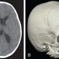
Optimal Time to Surgery in Patients With Traumatic Orbital Fracture ( Chapter 1.9 )
Clinical Question:
What is the optimal time to surgery in patients with traumatic orbital fracture?
Author Recommendation:
In children and adults with orbital fractures in order to achieve a complete recovery and prevent postsurgical enophthalmos and residual diplopia, clinicians should not delay indicated surgical repair.
What Does the Evidence Conclude?
| Quality of Evidence a | Strength of Recommendations b | Evidence |
|---|---|---|
| Very low | Strong | Evidence suggests that a delay in surgical repair is associated with poor clinical outcomes in most cases. The data is insufficient to determine the optimal surgical timing. Expert opinion is that immediate, within 24 hours, surgical repair should be recommended for younger patients and patients with nonresolving oculocardiac reflex, blow-out orbital fractures, and early enophthalmos. A poor prognosis of pediatric orbital fractures with entrapment resulting in a rapid development of muscle fibrosis and persistent diplopia may justify early surgical interventions in children with orbital fractures. |
a Quality of Evidence scale (GRADE): high, moderate, low, and very low. For more information on the GRADE rating system, see http://gdt.guidelinedevelopment.org/app/handbook/handbook.html
b The Guideline Elements Model: http://gem.med.yale.edu.easyaccess1.lib.cuhk.edu.hk/default.htm .
What Are the Parameters of Our Evidence Search?
| PICO | |
|---|---|
| Population | Children or adults with orbital fractures due to facial trauma Patient demographics Presence or absence of entrapment |
| Intervention | Shorter timing between the trauma and surgical intervention |
| Comparator | Longer timing between the trauma and surgical intervention |
| Primary outcome(s) | All-cause mortality, quality of life, function Treatment utilization All harms, including surgical complications |
Basis for and Determinants of the Strength of Recommendations:
Population:
Adults with orbital fractures
Setting:
Inpatient
Intervention:
Shorter timing between the trauma and surgical intervention
Comparator:
Delay in timing between the trauma and surgical intervention
| Outcomes | Risk With Intervention Per 1000 | Risk With Comparator Per 1000 | Relative Measure of Association (95% Confidence Interval) | Number of Participants (Studies) | Quality (GRADE) | Comments |
|---|---|---|---|---|---|---|
| Surgery After <7 Days Versus >7 Days After Trauma | ||||||
| Complete resolution of diplopia | 0 | 200 | OR 0.3 (0.01;8.46) | 10 (1 observational study) | Very low | No difference |
| Surgery After <2 Weeks Versus 2 Weeks to 6 Months After Trauma | ||||||
| Complete recovery | 730 | 250 | OR 6.9 (1.35;35.06) | 29 (1 observational study) | Very low | Favors shorter timing |
| Surgery After <2 Weeks Versus >2 Weeks After Trauma in Patients With Blow-Out Fracture | ||||||
| Diplopia | 63 | 149 | Peto OR 0.3 (0.1;0.9) | 442 cases | Very low | Favors shorter timing |
| Enophthalmos | 33 | 81 | Peto OR 0.2 (0.1;0.9) | 442 cases | Very low | Favors shorter timing |
| Surgery After <30 Days Versus >30 Days After Trauma | ||||||
| Complete resolution of preoperative motility restriction | 600 | 0 | OR 24.6 (1.30;462.34) | 40 (1 observational study of children) | Very low | Favors shorter timing |
| Complete resolution of diplopia | 516 | 0 | OR 19.2 (1.02;360.51) | 40 (1 observational study of children) | Very low | Favors shorter timing |
| Surgery After <24 Hours Versus >24 Hours After Trauma | ||||||
| Residual diplopia | 83 | 375 | OR 0.2 (0.01;1.84) | 20 (1 observational study of children) | Very low | No difference |
| Surgery After <4 Weeks Versus >4 Weeks After Trauma | ||||||
| Enophthalmos postoperative | NR | NR | P < .05 | 22 (1 RCT) | Very low | Favors shorter timing |
| Surgery After <8 Weeks Versus >8 Weeks After Trauma | ||||||
| Enophthalmos postoperative | 70 | 500 | OR 0.1 (0.01;0.46) | 51 (1 observational study) | Very low | Favors shorter timing |
Guideline Recommendations:
American Association of Oral and Maxillofacial Surgeons. Clinical Practice Guidelines for Oral and Maxillofacial Surgery, 2012. (AGREE II Score: Unavailable)
- •
This guideline considers time elapsed since injury as a risk factor for poor outcome but does not specify the optimal time for the surgery of orbital fractures.
Author Commentary:
Background
Orbital fractures are common and result in patients suffering with enophthalmos (displacement of the eyeball within the orbit), diplopia, orbital soft tissue entrapment, and nerve injury. There is no consensus around optimal timing for surgical repair of the fractured orbital floor. We systematically reviewed benefits and harms from early versus delayed surgery for orbital fractures.
Methods
We conducted a comprehensive search in PubMed, EMBASE, the Cochrane Library, Elsevier text mining tool database including National Institutes of Health RePORTER grant database, and clinicaltrials.gov trial registry up to June 2016 and did a hand-search of the reference lists. We included all studies that examined timing of surgical treatments for orbital fractures.
Results
We identified one clinical and two systematic reviews, 1 RCT, and 16 nonrandomized studies. The RCT was designed to examine treatment options and provide post hoc analysis of the association between timing and patient outcomes. A very small observational case series compared different timing and failed to provide reproducible data and to adjust for patient characteristics, surgical techniques, or the quality of the provided care. Adult case series analyzed various orbital fractures. Pediatric case series analyzed mostly blow-out orbital fractures.
Very low-quality evidence suggests that a delay in surgical repair is associated with poor clinical outcomes in most case series ( Table 1 ). The data is insufficient to determine the optimal surgical timing. A clinical review speculated that immediate, within 24 hours, surgical repair should be recommended for younger patients and patients with nonresolving oculocardiac reflex, blow-out orbital fractures, and early enophthalmos ( Appendix ). Studies did not stratify outcome rates by baseline entrapment. However, pediatric case series acknowledge a poor sensitivity in diagnosis of entrapment by radiologists and poor prognosis of pediatric orbital fracture with entrapment, resulting in a rapid development of muscle fibrosis and persistent diplopia.
Limitations
We downgraded the quality of evidence due to risk of bias in the body of evidence and heterogeneity and imprecision in treatment effects from small observational studies. We could not meta-analyze the data from case series due to differences in timing of surgery and definition of patient outcomes. The exact reason for surgical delay was not reported in the studies.
A single guideline defines a delay in surgical treatment after injury as a risk factor for poor outcome after any facial trauma but does not specify the optimal time for the surgery of orbital fractures. Future research is needed to determine the optimal timing for surgical repair of orbital fractures in patient subpopulations.
Conclusions
In children and adults with orbital fractures, in order to achieve a complete recovery and prevent postsurgical enophthalmos and residual diplopia, clinicians should not delay indicated surgical repair.
| Optimal Timing | Computed Tomography Findings |
|---|---|
| Immediate Within 24 hours | Diplopia present with computed tomography (CT) evidence of an entrapped muscle or periorbital tissue associated with a nonresolving oculocardiac reflex: bradycardia, heart block, nausea, vomiting, or syncope “White-eyed blow-out fracture.” Young patients (<18 years), history of periocular trauma, little ecchymosis or edema (white eye), marked extraocular motility vertical restriction, and CT examination revealing an orbital floor fracture with entrapped muscle or perimuscular soft tissue Early enophthalmos/hypoglobus causing facial asymmetry Significant globe displacement vision-threatening emergency |
| Within 2 weeks | Symptomatic diplopia with positive forced ductions, evidence of an entrapped muscle or perimuscular soft tissue on CT examination, and minimal clinical improvement over time Large floor fracture causing latent enophthalmos Significant hypo-ophthalmos Progressive infraorbital hypesthesia |
| Observation | Minimal diplopia (not in primary or downgaze), good ocular motility, and no significant enophthalmos or hypo-ophthalmos |
Glossary:
AGREE II, Appraisal of Guidelines for Research and Evaluation; CI, confidence interval; CT, computed tomography; GRADE, Grading of Recommendations Assessment, Development and Evaluation; NR, not reported; OR, odds ratio; RCT, randomized controlled trial; RePORTER, National Institutes of Health Research Portfolio Online Reporting Tools https://federalreporter-nih-gov.easyaccess1.lib.cuhk.edu.hk/ .
Recommended Citation:
Dorafshar A, Shamliyan TA; Elsevier Evidence-based Medicine Center: Evidence Report: Optimal time to surgery in patients with traumatic orbital fracture. Elsevier Evidence-Based Medicine Center. March 1, 2017.
Role of Surgery Versus Medical Treatment for Traumatic Optic Nerve Compression ( Chapter 1.9 )
Clinical Question:
What is the comparative effectiveness of surgery versus medical treatment for traumatic optic nerve compression?
Author Recommendation:
In children and adults with traumatic optic nerve compression, in order to achieve some improvement in visual acuity, clinicians should recommend systemic corticosteroids and consider rescue surgery in individual patients who do not improve after systemic corticosteroids.
What Does the Evidence Conclude?
| Quality of Evidence a | Strength of Recommendations a | Evidence |
|---|---|---|
| Low | Weak | Evidence suggests that systemic corticosteroids increase the rates of improved visual acuity when compared with observation in adolescents and adults with traumatic nonpenetrating optic nerve compression. Evidence suggests that there are no differences in the rates of improved visual acuity between methylprednisolone versus placebo, between dexamethasone versus methylprednisolone, or between megadoses versus high doses of methylprednisolone. |
| Very low | Weak | Evidence suggests that surgical optic nerve canal decompression alone or in combination with systemic corticosteroids increases the rates of improved visual acuity when compared with observation in adolescents and adults with traumatic nonpenetrating optic nerve compression. However, surgical optic nerve canal decompression is not better than systemic corticosteroids in improving visual acuity. Surgical optic nerve canal decompression combined with systemic corticosteroids is not better in improving visual acuity than systemic corticosteroids alone or surgery alone. |
a Quality of Evidence scale (GRADE): high, moderate, low, and very low. For more information on the GRADE rating system, see http://gdt.guidelinedevelopment.org/app/handbook/handbook.html .
b The Guideline Elements Model: http://gem.med.yale.edu.easyaccess1.lib.cuhk.edu.hk/default.htm .
What Are the Parameters of Our Evidence Search?
Basis for and Determinants of the Strength of Recommendations:
Population:
Adolescents or adults with traumatic nonpenetrating optic nerve compression
Setting:
Inpatient
Intervention:
Systemic steroids within 7–14 days after injury
Comparator:
Placebo, no active treatment, or active comparators
| Outcomes Comparisons | Risk With Intervention Per 1000 | Risk With Comparator Per 1000 | Relative Measure of Association (95% Confidence Interval) | Number of Participants (Studies) | Quality (GRADE) | Comments |
|---|---|---|---|---|---|---|
| Systemic Methylprednisolone Versus Placebo | ||||||
| Recovery of visual acuity defined as a decrease of at least 0.4 log MAR in visual acuity, 3 months | 688 | 533 | RR 1.3 (0.7;2.3) | 31 (1 RCT) | Very low | No difference |
| Systemic Steroids Versus Observation | ||||||
| Improvement in visual acuity | 449 | 346 Attributable events per 1000 treated 282 (112;453) | Peto OR 2.7 (1.5;4.9) NNT 4 (2;9) | 302 cases | Low | Favors systemic steroids |
| Visual acuity improved (log MAR) | NR | NR | MD 37.3 (20.7;53.9) SMD 0.8 (0.5;1.2) | 113 cases | Very low | Favors systemic steroids |
| High-Dose Dexamethasone Versus Megadose of Methylprednisolone | ||||||
| Visual improvement >2 lines, 2–17 months | 441 | 387 | RR 1.4 (0.3;5.8) | 65 (2 RCTs) | Very low | No difference |
| >3 lines improvement in best corrected visual acuities, 2 months Subgroup: No light perception at baseline | 400 | 400 | RR 1.0 (0.2;4.6) | 10 (1 RCT) | Very low | No difference |
| >3 lines improvement in best corrected visual acuities, 2 months Subgroup: preserved finger counts at baseline | 1000 | 1000 | RR undetermined | 9 (1 RCT) | Very low | No difference |
| Megadose Versus High Dose of Methylprednisolone | ||||||
| >1 lines improvement in visual acuity | 923 | 875 | OR 1.7 (0.1;31.9) | 21 cases | Very low | No difference |
| Early (Within 24 Hours From Trauma) Versus Delayed (Within 7 Days From Trauma) Treatment With Prednisolone 20 mg/kg Body Weight/Day for 3 Days in Children | ||||||
| >1 line improvement in visual acuity | 800 | 591 | Peto OR 2.8 (0.3;29.0) | 27 cases | Very low | No difference |
Population:
Adolescents or adults with traumatic nonpenetrating optic neuropathy
Setting:
Inpatient
Intervention:
Surgical optic nerve canal decompression
Comparator:
Systemic Corticosteroids or Observation
| Outcomes Comparison | Risk With Intervention Per 1000 | Risk With Comparator Per 1000 | Relative Measure of Association (95% Confidence Interval) | Number of Participants (Studies) | Quality (GRADE) | Comments |
|---|---|---|---|---|---|---|
| Surgical Optic Nerve Canal Decompression Versus Observation | ||||||
| Improvement in visual acuity | 397 | 105 | Peto OR 4.1 (1.4;12.6) | 77 cases | Very low | Favors surgery |
| Visual acuity improved (log MAR) | NR | NR | MD 26.0 (9.5;42.5) SMD 0.6 (0.2;1.0) | 117 cases | Very low | Favors surgery |
| Surgical Optic Nerve Canal Decompression Versus Systemic Corticosteroids | ||||||
| Improvement in visual acuity | 558 | 404 | Peto OR 1.4 (0.9;2.1) | 468 cases | Very low | No difference |
| Visual acuity improved (log MAR) | NR | NR | MD −11.3 (−25.9;3.3) SMD −0.3 (−0.6;0.1) | 132 cases | Very low | No difference |
| Surgical Optic Nerve Canal Decompression Plus Systemic Corticosteroids Versus Observation | ||||||
| Visual acuity improved (log MAR) | NR | NR | MD 20.9 (4.2;37.6) SMD 0.5 (0.1;0.8) | 112 cases | Very low | Favors surgery plus systemic corticosteroids |
| Surgical Optic Nerve Canal Decompression Plus Systemic Corticosteroids Versus Surgery Alone | ||||||
| Improvement in visual acuity, any | 286 | 533 | OR 0.4 (0.1;1.4) | 36 cases | Very low | No difference |
| Surgical Optic Nerve Canal Decompression Plus Systemic Corticosteroids Versus Systemic Corticosteroids Alone | ||||||
| Improvement in visual acuity | 313 | 471 | Peto OR 0.5 (0.2;1.2) | 100 cases | Very low | No difference |
| Surgical Optic Nerve Canal Decompression Plus Systemic Corticosteroids Within 7 Versus >7 Days After Injury | ||||||
| >3 lines improvement in visual acuity | 696 | 238 Attributable events per 1000 treated 458 (196;719) | Peto OR 6.0 (1.9;19.4) NNT 2 (1;5) | 44 cases | Very low | Favors early treatment |
| Early (Within 7 Days) Versus Late (>7 Days After Injury) Endoscopic Optic Nerve Decompression and Postoperative Corticosteroid in Children | ||||||
| >3 lines improvement in visual acuity | 964 | 462 Attributable events per 1000 treated 503 (223;782) | OR 31.5 (3.2;306.2) NNT 2 (1;4) | 41 cases | Very low | Favors early treatment |
Guideline Recommendations:
American Association of Oral and Maxillofacial Surgeons. Clinical Practice Guidelines for Oral and Maxillofacial Surgery, 2012. (AGREE II Score: Unavailable)
This guideline lists the following procedures for the management of orbital injuries but does not address preferred treatments for optic nerve compression:
- A.
Observation based on limited severity of fracture, displacement, and mobility
- B.
Open treatment (including endoscopically assisted and computed tomography-guided navigation)
- C.
Orbital reconstruction
- D.
Medial and/or lateral canthopexy
- E.
Nasolacrimal reconstruction
- F.
Antimicrobials as indicated
- G.
Control of pain
- H.
Drains for management of dead spaces or contaminated wounds when judgment dictates
- I.
Instructions for posttreatment care and follow-up
Author Commentary:
Background
Treatment options for traumatic optic nerve compression include observation, large dose of systemic corticosteroids, or surgical treatments, with no consensus on which option is superior to the others in preventing devastating blindness. The lack of direct randomized trials results in variable usual practices among hospitals and clinicians. We systematically reviewed the comparative effectiveness of surgical and medical treatment options for children and adults with traumatic optic nerve compression.
Methods
We conducted a comprehensive search in PubMed, EMBASE, the Cochrane Library, Elsevier text mining tool database including National Institutes of Health RePORTER Grant database, and clinicaltrials.gov trial registry up to August 2016 and did a hand-search of the reference lists. We used random effects models to pool relative risk from RCTs, Peto odds ratios from sparse controlled nonrandomized studies, and arcsine transformed crude rates of the outcomes from uncontrolled case series and case reports.
Results
We identified 4 meta-analyses, 1 systematic review, 3 RCTs, 1 controlled International Optic Nerve Trauma Study, and 42 publications of case series. Three Cochrane reviews did not provide quantitative data. Primary studies included children and adults and rarely reported results by age groups. The exact treatment regimens and outcomes measurements varied across individual reports. Primary studies of surgical treatments inconsistently reported concomitant use of pre- and postoperative systemic steroids. Many retrospective case series reported outcomes in patients who received surgery after systemic corticosteroids failed to improve visual acuity. Surgical techniques included extra- or intracranial, orbital, transethmoidal, or endoscopically assisted transconjunctival approaches. Studies did not compare different surgical techniques and did not explain the selection of the techniques used depending on patient characteristics.
Low-quality evidence suggests that systemic corticosteroids increase the rates of improved visual acuity when compared with observation in adolescents and adults with traumatic nonpenetrating optic nerve compression ( Table 1 ). Very low-quality evidence suggests that there are no differences in the rates of improved visual acuity between methylprednisolone versus placebo, between dexamethasone versus methylprednisolone, or between megadoses versus high doses of methylprednisolone ( Table 1 ).
Early (within 24 hours from trauma) versus delayed (within 7 days from trauma) prednisolone administration does not result in greater rates of improved visual acuity in children with traumatic nonpenetrating optic nerve compression ( Table 1 ).
Very low-quality evidence suggests that surgical optic nerve canal decompression alone or in combination with systemic corticosteroids increases the rates of improved visual acuity when compared with observation in adolescents and adults with traumatic nonpenetrating optic nerve compression ( Table 2 ). However, surgical optic nerve canal decompression is not better than systemic corticosteroids in improving visual acuity ( Table 2 ). Surgical optic nerve canal decompression combined with systemic corticosteroids is not better in improving visual acuity than systemic corticosteroids alone or surgery alone ( Table 2 ). Early surgery results in greater rates of improvement in visual acuity than later surgery in adults and children ( Table 2 ).
Indirect comparison of uncontrolled case series suggests that the rates of the improved visual acuity are slightly lower after observation and are very similar after systemic corticosteroids alone or after surgery in patients who did not experience improved vision after systemic corticosteroids ( Appendix Table 1 ). Uncontrolled case series suggest that patients with optic canal fracture and patients without baseline light perception have poor prognosis. The rates varied substantially among single institutions, probably because of differences in hospital protocols regarding indication for steroids or surgery, and probably due to differences in the quality of provided care.
Limitations
We downgraded the quality of evidence due to high risk of bias in the body of evidence and heterogeneity and imprecision in treatment effects from small studies. The evidence regarding comparative effects from various steroids and surgical techniques on complete vision restoration, mortality, quality of life, and treatment utilization was insufficient. Small studies attempted to examine correlation between baseline vision loss and treatment outcomes but failed to identify valid evidence for individualized treatment decisions. Authors came to the somewhat obvious conclusion that patients with minimal baseline vision loss have better vision after the treatments. Studies did not report outcomes in patients with different demographics or comorbidities.
We found one guideline that describes treatment options for orbital injuries but does not address preferred treatments for indirect optic nerve compression. Future research is needed to examine the comparative effectiveness and safety of surgical versus medical treatments in patients with traumatic optic nerve compression and various demographic groups and comorbidities.
Conclusions
In children and adults with traumatic optic nerve compression, in order to achieve some improvement in visual acuity, clinicians should recommend systemic corticosteroids and consider rescue surgery in individual patients who do not improve after systemic corticosteroids.
| Treatments | % With Improvement (95% CI) * | Cases |
|---|---|---|
| Conservative treatment | 32.8 (23.6;45.5) | 168 cases |
| Corticosteroids | 54.9 (44.8;67.2) | 250 cases |
| Surgery | 54.9 (45.9;65.6) | 304 cases |
| Surgery + steroid | 35.0 (21.1;58.2) | 61 cases |
| Surgery in patients who did not improve after systemic corticosteroids | 57.8 (48.9;68.2) | 1445 cases |
Glossary:
AGREE II, Appraisal of Guidelines for Research and Evaluation; CI, confidence interval; GRADE, Grading of Recommendations Assessment, Development and Evaluation; log MAR, log of the minimum angle of resolution; MD, mean difference; NNT, number needed to treat to achieve an outcome in 1 patient; NR, not reported; OR, odds ratio; RCT, randomized controlled trial; RePORTER, National Institutes of Health Research Portfolio Online Reporting Tools https://federalreporter-nih-gov.easyaccess1.lib.cuhk.edu.hk/ ; RR, relative risk.
Recommended Citation:
Dorafshar AH, Davidson E, Reddy S, et al. Role of surgery versus medical treatment for traumatic optic nerve compression. Elsevier Evidence-Based Medicine Center. March 1, 2017.
Fixation Points for Traumatic Zygomaticomaxillary Complex Fractures ( Chapter 1.12 )
Clinical Question:
What are the benefits and harms of various point fixation techniques in patients with traumatic zygomaticomaxillary complex fractures?
Author Recommendation:
In adults with isolated, laterally displaced zygomatic bone fractures, in order to achieve stable fixation and reduce vertical dystopia, clinicians should use 3-point over 2-point fixation with miniplates.
What Does the Evidence Conclude?
| Quality of Evidence a | Strength of Recommendations b | Evidence |
|---|---|---|
| Very low | Weak | Evidence suggests that 3-point fixation results in higher rates of stable fracture fixation and smaller vertical dystopia when compared with 2-point fixation in adults with isolated, laterally displaced zygomatic bone fractures. |
| Very low | Weak | There are no differences in patient outcomes between single-point fixation with titanium miniplate versus single-point fixation with bioresorbable plates or between 1-point fixation with titanium miniplate versus fixation with Kirschner wire in adults with zygomatic complex fracture. |
| Insufficient | Weak | Uncontrolled case series suggest that the rates of beneficial treatment outcomes including patient satisfaction with postoperative appearance, facial symmetry, or stable fracture are more than 90% across various fixation points, while the rates of postsurgical complications and infections vary across institutions regardless of the number of fixation points. 4-point fixation results in bony union and facial symmetry in patients with isolated zygomatic arch fracture and insufficient intraoperative reduction after closed reduction and a single-fixation point. |
Related posts:


Stay updated, free articles. Join our Telegram channel

Full access? Get Clinical Tree


