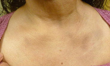
Figure 15.1
Erythema dyschromicum perstans. Ash-like, slate gray to blue, oval macules and patches are noted on the chest (a) and antecubital fossa (b)
Differential Diagnosis
The differential diagnosis of this disorder is broad. The most frequent cause of confusion and controversy is with lichen planus pigmentosus, which is characterized by hyperpigmented dark-brown macules with no active border and a noncharacteristic distribution that predominates in sun exposed areas and flexor folds. Its course is characterized by exacerbations and remissions and occasionally accompanied by pruritus. Lichenoid drug eruptions often involve trunk and limbs symmetrically and are photodistributed. Atrophic lichen planus can occur anywhere, but favors the flexor wrists, trunk, medial thighs, shins, dorsal hands, and lower back. Postinflammatory hyperpigmentation is characterized by brown discoloration. Idiopathic eruptive macular pigmentation is characterized by asymptomatic, hyperpigmented pigmented macules involving the neck, trunk, and proximal extremities. Pityriasis rosea typically begins with a solitary patch, and then progresses to a generalized exanthem of bilateral and symmetric macules with a collarette of scale oriented within the long axes along cleavage lines. Patients with hemochromatosis present with diffuse hyperpigmentation, but usually have cirrhosis and diabetes mellitus. Macular urticarial pigmentosum presents with widespread yellow tan to red brown macules and usually involves the trunk more than the extremities. Treponemal eruptions, such as secondary syphilis, may also resemble erythema dyschromicum perstans. Therefore, serological testing for treponemes is appropriate when encountering this type of outbreak (Bahadir et al. 2004).
Histopathology
A punch biopsy was performed on the border of an active macule on the antecubital fossa. Hyperkeratosis, a thinned epidermis, focal vacuolar changes in the basal layer of the epidermis, superficial perivascular lymphohistiocytic infiltration, and pigment incontinence were present (Elder 2009).
Laboratory Studies
A complete blood count and comprehensive metabolic panel were normal. A human immunodeficiency virus test and rapid plasma reagin test were negative.
Diagnosis
Erythema dyschromicum perstans (EDP)
Case Treatment
The diagnosis of the disorder was discussed with the patient, along with its difficulty in treatment. Treatment options were discussed including hydroquinone, clofazimine, dapsone, and phototherapy. The patient declined clofazimine and dapsone due to the side effects of the medications. She was started on hydroquinone 4 % cream twice daily to affected areas. She also underwent narrowband UVB phototherapy three times a week. A broad spectrum sunscreen with an SPF of 30 was recommended for use daily. At 2 month follow-up, she noted mild improvement.
Discussion
Erythema dyschromicum perstans (EDP) or “ashy dermatosis” was first described in 1957 by Ramirez in Salvadorans as an “asymptomatic, ash-colored, macular hyperpigmentation that is slowly progressive and leaves a permanent discoloration” (Ramirez 1957). EDP is characterized by ash-like, slate gray, oval macules and patches with erythematous borders that range in diameter from 0.5 cm to very large confluent patches. Individual lesions can be oval, irregular, or polycyclic in shape and continue to grow slowly. Lesions initially appear on the trunk and spread centrifugally to the extremities (Figs. 15.1 and 15.2). The scalp, palms, soles, and mucous membranes are rarely affected. As the lesions extend, the erythematous border eventually disappears within several months, so it may no longer be evident when the patient is examined. EDP has traditionally been characterized by its poor response to medication and its tendency to persist indefinitely in adult patients (Chun et al. 2009).


Figure 15.2
Erythema dyschromicum perstans. Ash-like, hyperpigmented patches are noted on the chest
Erythema dyschromicum perstans usually occurs in the second or third decade of life and is most common in individuals from Central and South America, although cases have been described from different parts of the world. It is somewhat more common in women. EDP usually appears in adults, but some isolated cases and a small series of eight cases have been reported in prepubertal children (Silverberg et al. 2003).
The cause of EDP is unclear. Many consider EDP to be a variant of lichen planus actinus (Naidorf and Cohen 1982). Infections, such as whipworm, HIV (Nelson et al. 1992), exposure to environmental toxins, such as pesticides, ammonium nitrate, cobalt, radiocontrast media, and fungicides, and medications, such as oral antibiotics and benzodiazepines, have all been implicated in the etiology of EDP (Silverberg et al. 2003). Systemic conditions such as thyroid disease and chronic hepatitis (Kontochristopoulos et al. 2001) have also been implicated.
Stay updated, free articles. Join our Telegram channel

Full access? Get Clinical Tree


