Abstract
Frontal sinus and maxillary sinus fractures involving the orbital floor have traditionally been repaired with open approaches. With advances in nasal endoscopy and endoscopic instrumentation, less invasive approaches to these fractures are now feasible. This chapter discusses the surgical anatomy, radiographic evaluation, perioperative care, and surgical techniques involved with the endoscopic repair of frontal sinus and orbital floor fractures.
Keywords
frontal sinus, maxillary sinus, fracture, endoscopic, surgical technique
Background
The paranasal sinuses are aerated spaces in the middle and upper third of the face that develop during the first and second decades of life in the frontal, ethmoid, maxillary, and sphenoid bones. The theoretical role of these structures in facial trauma is to provide shock absorption and dispersal of forces in order to prevent more severe injuries to critical anatomical structures such as the brain and eyes.
The frontal sinuses, located in the frontal bone, are paired cavities that are present at birth in only 12% of neonates. These sinuses typically form their adult shape by age 12, and continue to fully aerate into early adulthood. Due to protection from thick cortical bone anteriorly, the frontal sinus is least likely to sustain a fracture, accounting for 5%–15% of all maxillofacial injuries. As a result, frontal sinus fractures typically result from high-impact mechanisms, such as motor vehicle collisions and assault. These fractures are typically classified by their location, and may involve the anterior table or outer wall of the frontal sinus, the posterior table or inner wall of the frontal sinus, or both. Disruption of the inferior portion of the frontal sinus may compromise the frontal sinus outflow tract or frontal recess. As a result, long-term sequelae of these fractures may include chronic frontal sinusitis, mucocele, mucopyocele, and intracranial complications.
The maxillary sinuses are paired cavities in the maxilla that are located inferior to the orbits bilaterally. They are the largest sinuses present at birth, and aerate in an inferolateral direction to extend laterally to the zygomatic recess and inferiorly to the level of the nasal floor by age 12. While the majority of maxillary sinus fractures are inconsequential and do not require surgical repair, the maxillary sinus roof is the orbital floor, and fractures in this region may result in orbital injury.
Traditional open surgical approaches to the mid- and upper face have been extensively described in the literature and provide good access to fractures in this region. Traditional approaches to the frontal sinus included osteoplastic flaps or sinus obliteration via a bicoronal incision as well as cranialization. Open approaches to the orbital floor included transconjunctival and transcaruncular approaches.
With advances in nasal endoscopy and endoscopic instrumentation, less invasive approaches to these fractures are now feasible. While these approaches may offer a less invasive means to manage frontal and orbital floor fractures, evidence to date is limited to case reports and case series. Moreover, long-term outcomes in these patients have yet to be established. Regardless, a growing body of evidence suggests that endoscopic approaches have acceptable safety and efficacy, and represent a promising means to manage these injuries with decreased morbidity and better cosmesis than traditional approaches. These endoscopic approaches will be discussed further in this chapter.
Surgical Anatomy
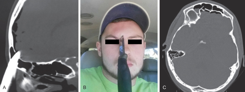
The frontal sinuses are paired sinuses present in the frontal bone separated by a variable intersinus septum. The anterior table of the frontal sinus is the bone just deep to the skin and subcutaneous tissue of the brow. The posterior table of the frontal sinus is the bone just anterior to the dura mater of the frontal lobe. The anterior table (4–12 mm) is significantly thicker than the posterior table (0.1–4.8 mm). The anatomy of the frontal sinus and frontal recess, which is shaped like an inverted funnel, is highly variable. The anteroposterior (A-P) distance of the frontal recess is perhaps the most important anatomical consideration, with a distance of at least 5 mm required in order to adequately instrument the area endoscopically. When considering endoscopic approaches it is important to understand that the frontal recess is bordered laterally by the lamina papyracea of the orbit, medially by the vertical lamella of the middle turbinate, and lateral lamella of the cribriform plate, anteriorly by the frontal beak, and posteriorly by the posterior table and anterior skull base. The configuration of the frontal recess is highly variable, and may be restricted by ethmoid pneumatization. Pneumatization anteriorly is typically by agger nasi or supra-agger nasi cells, which may extend into the frontal sinus itself. Pneumatization posteriorly is typically by air cells superior to the bulla ethmoidalis, or suprabullar cells. These suprabullar cells may also extend superiorly into the frontal sinus and laterally above the orbit. Finally, frontal septal cells are anterior ethmoid cells that pneumatize medially and may be contiguous with the frontal sinus septum, pushing the frontal drainage pathway laterally. These cells may also be described as types 1–4 frontal cells. A computed tomography (CT) of the sinuses showing frontal recess anatomy is provided ( Fig. 1.8.1 ).
The maxillary sinus is located just lateral to the nasal cavity and, most importantly relative to facial trauma, shares its superior border with the orbital floor. Endoscopically, the maxillary sinus is approached via the osteomeatal complex, which is generally well visualized by gentle medialization of the middle turbinate endonasally. The key anatomical structures when approaching the maxillary sinus include the maxillary line, which is the junction of the hard maxillary bone and the lacrimal bone, and the uncinate process. The uncinate process is a thin, sickle-shaped bone that is typically removed at the beginning of any approach to the maxillary sinus in order to visualize the natural ostium of the sinus. This ostium must be identified and widened in continuity with the maxillary antrostomy in order to prevent mucociliary recirculation, which may lead to chronic maxillary sinusitis.
Clinical Presentation
In a large retrospective cohort study, frontal sinus fractures most commonly occurred in males, with an average age of 30 years. Fractures most commonly involved both the anterior and posterior tables, followed by the anterior table alone. Only 3% were isolated posterior table fractures, and only 2% involved the frontal recess alone. As frontal fractures are associated with high-impact mechanisms of injury, clinical evaluation must consider other associated fractures, injuries to the skull base, and intracranial injuries. Physical examination should begin with a primary trauma survey. In patients with frontal sinus fractures, focused physical examination should include any contour deformity of the glabella and brow, and a cranial nerve examination. Forehead paresthesias may be present if fractures involve the supraorbital and/or supratrochlear nerves. Any lacerations should be noted, and may be considered as potential ports for direct surgical approaches. Anterior rhinoscopy should be performed as an overview assessment for any nasal septal deviation, injury, or hematoma. A more comprehensive nasal endoscopic examination should be offered to assess for cerebrospinal fluid (CSF) leak, particularly in patients with known posterior table injury. In these cases, it is important to obtain consultation with a neurosurgeon for operative planning.
Orbital floor fractures are typically due to trauma to the globe and periorbita, with common mechanisms of injury being motor vehicle accidents and personal assaults. Assessment of patients with orbital floor fractures after primary trauma survey should include a thorough evaluation of the eye and orbit. Physical examination should include gross visual fields and assessment of extraocular motion. Malar paresthesias are common in these patients. They result from injury to the infraorbital nerve as it travels through the infraorbital groove and foramen, which is a weak spot in the orbital floor and a common area for fractures to occur. The patient should also be assessed for enophthalmos, although this typically develops in a delayed fashion. Cosmetic deficits such as contour deformity of the midface or midface instability, as well as the possibility of dental involvement, should also be considered. An ophthalmology consultation should be considered where available in all patients suffering an orbital fracture.
Radiological Evaluation
The maxillofacial CT scan is the gold standard for the radiological assessment of any maxillofacial fracture. This scan should be performed with thin slices, no greater than 1 mm spacing, to achieve the appropriate resolution for assessment of paranasal sinus fractures. Bone windows are important to determine the location and extent of fractures, and soft tissue windows should also be examined to assess for injury to the surrounding soft tissue structures.
Similar to all fractures, frontal sinus fractures should be examined in the axial, coronal, and sagittal planes. Of these planes, the axial and sagittal provide the most important information. These planes allow the examiner to determine the location of the fracture with respect to the anterior and posterior tables as well as any potential for the fracture to occlude the frontal recess. Displacement and comminution of the frontal bones may also be assessed in these planes and have a significant impact on management. For instance, nondisplaced, noncomminuted fractures may be managed expectantly, particularly when isolated to the anterior table. Conversely, more displaced, comminuted fractures and those that involve the posterior table are typically managed surgically.
Maxillary sinus fractures are also assessed using a maxillofacial CT scan. These fractures must be visualized in all three planes and location of the fractures with respect to the orbit is of the utmost importance. As with frontal sinus fractures, the degree of displacement and comminution also play a role in management. Orbital floor fractures are typically repaired either urgently in cases of orbital entrapment, or electively in larger fractures that may result in enophthalmos.
Surgical Indications
Absolute indications for repair of frontal sinus fractures include overt evidence of CSF leak or involvement of the frontal recess with concern for future frontal sinus outflow obstruction that could lead to chronic frontal sinusitis and/or mucocele formation. Whereas fractures in patients presenting with CSF leak should be repaired within 48 hours to reduce the risk of complications such as pneumocephalus and meningitis, fractures involving the frontal recess without evidence of intracranial involvement may be repaired on an elective basis. A relative indication for repair of frontal sinus fractures is contour deformity of the glabella and the resultant cosmetic defect. These repairs are typically performed once acute edema has resolved to obtain an appropriate cosmetic result.
Indications for repair of maxillary sinus fractures include orbital floor fractures with evidence of entrapment and/or those involving greater than 50% of the orbital floor. Such fractures have the potential to cause long-term sequelae such as ophthalmoplegia, diplopia, and visual deficits. Cosmetic deformity, such as enophthalmos, is also a concern in these cases. While surgical repair of these fractures is indicated, the timing of repair remains controversial and ranges from emergent repair to outpatient repair on an elective basis.
Transnasal Endoscopic Approach to Frontal Sinus Fractures
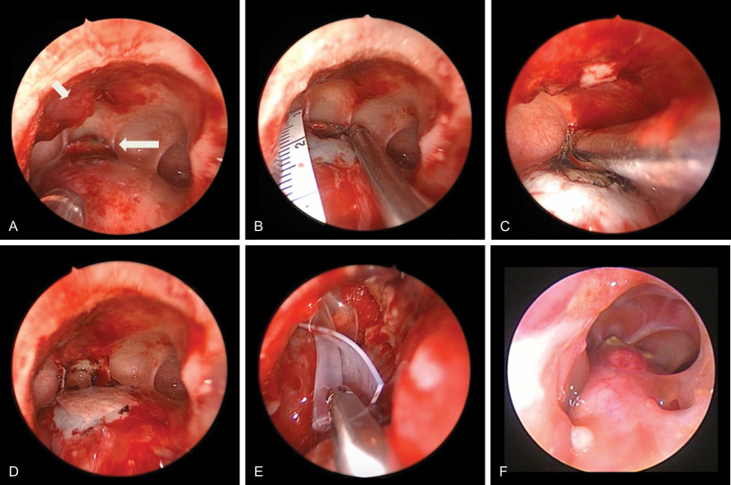
While endoscopic-assisted approaches to both the anterior and posterior tables have been described in the literature, the transnasal endoscopic approach will be described here ( Figs. 1.8.2 and 1.8.3 ). Endoscopic reduction of anterior table fractures should be performed within 10 days before significant healing to avoid difficulty in closed reduction of the segments. Operative intervention for posterior table injuries depends on the extent of the fractures and presence of CSF leak. Significant injuries should be addressed in a timely fashion (also usually within 10 days) when the patient’s condition is stable for anesthesia. While small linear fractures with CSF leak could be managed conservatively, it should be noted CSF leaks can present years after conservative management or previous open procedures.
Related posts:
 Assessment of the Patient With Traumatic Facial Injury
 Frontal Bone and Frontal Sinus Injuries
 Le Fort Fractures
 Secondary Reconstruction of Facial Soft Tissue Injury and Defects
 Geriatric and Edentulous Maxillary and Mandibular Fractures
 Secondary Microvascular Reconstruction of the Traumatic Facial Injury
Stay updated, free articles. Join our Telegram channel

Full access? Get Clinical Tree


