This article focuses on the contribution of acellular dermal matrices (ADMs) to immediate breast reconstruction. The current literature on ADMs is reviewed and the potential advantages and disadvantages of their use are highlighted. Technical considerations on how to effectively use these materials is presented.
- •
Immediate breast reconstruction is a safe and reliable approach to breast reconstruction.
- •
Use of acellular dermal matrices (ADMs) does not necessarily increase the rate of tissue expansion or final fill volumes after immediate 2-stage breast reconstruction.
- •
ADMs are associated with an increased complication rate in breast reconstruction compared with traditional techniques.
- •
ADMs can be used in the setting of the irradiated breast.
- •
ADMs should be used judiciously in immediate breast reconstruction in the following situations: (1) eliminate dead space after nipple-sparing mastectomy; (2) insertion of the pectoralis major muscle is high, prohibiting complete coverage of the inferior and lateral aspects of the tissue expander with serratus fascia and muscle.
- •
ADMs may have an important role in direct-to-implant single-stage breast reconstruction.
Overview of immediate versus delayed breast reconstruction
Higher complication rates in immediate than in delayed breast reconstruction are reported in the literature. Moreover, in many practice settings, coordinating immediate reconstruction with the breast surgery service and a busy operative schedule can be challenging. As a result, some practitioners are reluctant to incorporate immediate breast reconstruction into their practices. However, numerous reports have demonstrated safe and reliable techniques for immediate breast reconstruction with prosthetic devices. This article focuses on the contribution of ADMs to immediate breast reconstruction.
The current literature on ADMs is reviewed and the potential advantages and disadvantages of their use are highlighted. Technical considerations on how to effectively use these materials is presented.
Immediate Breast Reconstruction is Safe and Reliable
Since skin-sparing mastectomy was shown to be an oncologically sound procedure in the 1990s, implant-based breast reconstruction has become commonplace. In 2010, 93,083 breast reconstruction procedures were performed. This number is an increase of 8% from 2009. Most patients underwent a 2-stage reconstruction with a tissue expander and implant (62,081). A significantly smaller number of single-stage, implant-only reconstructions (9452) were performed. In the years ahead, the number of women who will undergo immediate reconstruction is likely to increase.
In a seminal article from 2002, Alderman and colleagues published one of the first outcome studies on postmastectomy breast reconstruction. The article reviewed the complication rates associated with autologous as well as implant-based reconstruction over a 2-year period in 326 patients. The study concluded that immediate breast reconstruction, regardless of the reconstructive technique, was associated with higher complication rates than delayed reconstruction. With respect to implant reconstruction, the study examined 65 patients who underwent immediate reconstruction and 14 who underwent delayed reconstruction. The rate of major complications was 46% and 21% respectively ( P = .089). Moreover, log regression analysis of major complications identified immediate reconstruction as an independent risk factor for developing a major complication with an odds ratio of 2.71. Many plastic surgeons continue to cite the report by Alderman and colleagues as reason not to pursue immediate breast reconstruction.
In the intervening years, numerous studies have shown that immediate breast reconstruction is a safe procedure with a low complication rate.
Cordeiro and colleagues reviewed a single surgeon’s 12-year experience with early postoperative complications in 1522 reconstructions in 1221 patients. All patients underwent immediate postmastectomy reconstruction with a tissue expander, subsequent expansion, and a secondary procedure to exchange the tissue expander for a permanent implant.
Complications included:
- •
Hematoma
- •
Mastectomy flap necrosis
- •
Seroma
- •
Infection.
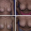
The observed overall complication rate was 5.8%. No increase in complications was noted in patients who underwent chemotherapy as part of their treatment regimen. However, the complication rate was higher in patients who had a history of chest wall irradiation. Review of the same cohort revealed that immediate 2-stage breast reconstruction yields good long-term aesthetic results with high patient satisfaction rates.
What is Acellularized Dermal Matrix?
Many different products constitute the acellularized dermal matrix genre of implantable medical devices. The most commonly used and studied is AlloDerm® (LifeCell Corp., Branchberg, NJ, USA). AlloDerm ® is cadaveric human dermis that has undergone a proprietary process to remove any antigenic properties of human skin. The end product is a biological matrix with the propensity to engraft into a vascularized tissue bed. It is not a sterile product, but it is aseptic, having been aggressively treated with a cocktail of antibiotic solutions to eliminate any organisms that may cause an infection. It comes in many different sizes and thicknesses, and has been described for numerous applications including:
- •
Abdominal wall reconstruction
- •
Static slings for facial reanimation
- •
Cleft palate repair
- •
Gynecologic and urologic reconstruction
- •
Breast reconstruction.
Other products include Strattice ™ (LifeCell Corporation, Branchburg, NJ, USA), an acellularized porcine dermis. Strattice ™ tends to be less elastic and resistant to deformational forces. One purported benefit of Strattice ™ compared with Permachol (Covidien, Mansfield, MA, USA) is that Strattice ™ is not cross-linked, theoretically allowing for more rapid neovascularization of the construct.
ADM s Emerge as a Popular Adjunct to Submuscular Immediate Implant Reconstruction
The first report of ADMs and breast surgery was for the treatment of contour abnormalities in cosmetic breast implant cases. Baxter in 2003 described a series of techniques using AlloDerm ® to correct difficult cosmetic problems such as synmastia, rippling, and malposition of implants. It was not until 2005, when Breuing and Warren reported the use of AlloDerm ® as an inferolateral sling in immediate breast reconstruction, that ADMs were identified as a useful tool in breast reconstruction. Breuing and Warren described a series of 10 patients in whom AlloDerm ® was used to augment a subpectoral pocket. The use of AlloDerm ® allowed for a 1-stage reconstruction with tight control of the degree of lower pole fullness. The potential advantage of decreasing the amount of time needed for tissue expansion and the notion of improved contour of the reconstructed breast mound was also introduced. Salzberg reported a similar technique used in 49 patients.
ADM s Have Many Perceived Benefits But Are Associated with Greater Complication Rates
Since these initial reports, the advantages of using ADMs have been highlighted in the literature and include decreased rates of capsular contracture and less postoperative pain; improved breast contour and camouflage of the implant; increased initial fill volume of tissue expanders in 2-stage reconstruction; 1-stage reconstruction with permanent implants.
Few of these claims are supported with prospective clinical studies. In addition, a randomized prospective study regarding the use of ADM and breast reconstruction does not exist. However, large retrospective cohort studies have provided some helpful observations. Preminger and colleagues performed a matched-cohort retrospective study to investigate whether the use of AlloDerm ® in immediate breast reconstruction affected the rate of tissue expansion or complications. Patients who underwent a 2-stage reconstruction with AlloDerm ® did not have a greater rate of expansion than patients who underwent a 2-stage breast reconstruction without AlloDerm ® . In addition, there was no significant difference in the rate of complications between the 2 groups. In this study, the investigators did not attempt to fill the expanders over aggressively in patients with or without ADM, which may explain the findings reported.
In a follow-up study, Antony and colleagues reviewed the Memorial Sloan-Kettering Cancer Center experience with ADM in 153 2-stage breast reconstructions. The study revealed that advanced age, increased body mass index, and axillary dissection are independent risk factors for developing a complication in ADM/tissue expander breast reconstruction. Specifically, the ADM group had higher rates of seroma (7.2%) and reconstructive failure (5.9%) compared with the non-ADM cohort. Most cases of reconstructive failure were attributed to infection (3.3%).
These findings are confirmed by Chun and colleagues in a review of 146 cases of ADM/implant-based reconstruction. Regression analysis showed that ADM use increased the risk of seroma formation more than 4 times and increased the risk of infection 5 times compared with a cohort who underwent non–ADM implant-based reconstruction. Although not statistically significant, Chun and colleagues noted that mastectomy skin flap necrosis occurred with greater frequency in the ADM group. This finding was attributed to higher initial fill volumes and a tendency to retain more mastectomy flap skin in ADM/implant-based reconstruction. When cases of skin flap necrosis were excluded, a statistically significant increase in postoperative seroma rate was observed. However, the noted increase in the rate of major postoperative complications did not reach statistical significance. Thus, Chun and colleagues concluded that seroma might predispose patients having ADM/implant-based breast reconstruction to infection.
ADM and Immediate Breast Reconstruction in the Irradiated Breast
In appropriately selected patients, neoadjuvant and adjuvant radiation therapy does not seem to preclude the use of ADM in immediate prosthetic breast reconstruction. Breuing and Colwell describe their experience with 5 patients who underwent immediate tissue expander implant or implant reconstruction with AlloDerm ® . All patients had adjuvant radiation therapy within 6 months of reconstruction and were followed for at least 2 years after radiation therapy was complete. None of the 5 patients developed capsular contracture or implant loss.
In a larger study, Spear and colleagues reviewed their experience with 58 patients who underwent ADM augmentation of the subpectoral pocket in 2-stage breast reconstruction. Compared with dual-plane and more traditional submuscular techniques, the use of ADM was associated with a higher rate of seroma and infection. However, radiation therapy was the only variable shown to increase the risk of developing an infection. Based on these data, Spear and colleagues concluded that a history of radiation therapy does not exclude patients from the use of ADM as part of their reconstructive regimen.
As stated previously, Antony and colleagues showed that ADM use in immediate breast reconstruction can result in an increased risk of postoperative complications. However, multivariate analysis failed to reveal neoadjuvant radiation as an independent risk factor for postoperative complications. The investigators cited careful patient selection as a possible explanation for this observation. Specifically, they reported that candidates for breast reconstruction with tissue expanders and ADMs must have no evidence of radiation damage to the mastectomy skin flaps and were at least 1-year status post completion of radiation therapy.
Two-Stage Versus 1-Stage Breast Reconstruction
An advantage of using ADMs in breast reconstruction is the option of immediate breast reconstruction with permanent implants. As discussed earlier, AlloDerm ® in this setting provides an extension of the subpectoral pocket and control over the aesthetic unit of the inferior pole.
One-stage breast reconstruction: Wise-pattern variation
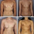
Derderian and colleagues describe a variation of Wise-pattern breast reconstruction that uses AlloDerm ® and a vascularized dermal pedicle to improve breast mound shape and protect against implant exposure at the T-point of the mastectomy flap closure. In their study of 20 patients, a 25% T-point breakdown rate was reported. All the wounds healed with local wound care. Their study describes an innovative use of ADM and redundant local tissue to improve a previously described technique for immediate reconstruction. A caveat is that the Wise-pattern technique is only appropriate for patients with large breasts, who tend to have redundant skin after parenchymal resection. Generally, successful single-stage, direct-to-implant reconstruction with ADM requires well-perfused, viable mastectomy skin flaps, and a reconstruction that is similar to, or smaller than, the original volume of the breast.
Two-stage breast reconstruction
The example mentioned earlier shows the usefulness of ADMs in a single-stage technique. However, there are no prospective randomized clinical studies that compare the outcomes of 2-stage tissue expander/implant versus direct implant techniques.
The typical technique for immediate prosthetic breast reconstruction at Memorial Sloan-Kettering Cancer Center is a 2-staged approach. Immediately after mastectomy, a tissue expander is placed in a subpectoral pocket. After a series of expansions in the outpatient clinic, the patient undergoes exchange of the tissue expander for a permanent implant. Use of ADM occurs at the time of mastectomy and expander placement to elongate the pectoralis as an inferolateral sling. During the second stage, capsulotomy, capsulorrhaphy, and contralateral symmetry procedures are performed as needed.
Patients who have already undergone mastectomy and present for delayed reconstruction with tissue expanders and implants typically do not require ADM at the time of expander placement or exchange to the permanent implant. If the mastectomy flaps have previously been irradiated, delayed reconstruction almost always requires autologous tissue, with or without a prosthetic device.
Indications for ADM Placement
In the senior author’s practice, ADM is used as an adjunct in immediate prosthetic breast reconstruction, not as a requirement. Most implant-based breast reconstructions can be performed using either total or partial muscular coverage of the expander, depending on the local anatomy and the quality of the mastectomy skin flaps. However, there is a role for ADM in prosthetic breast reconstruction to extend the length of the pectoralis major muscle when its insertion is high relative to the inframammary fold, particularly if the anterior rectus sheath has been violated during the mastectomy. In this situation, the distance between the inferior border of the pectoralis major muscle, anterior rectus sheath, and serratus fascia and muscle either prohibits complete muscular coverage of the expander or creates an excessively tight lower pole that is likely to result in a high-riding, taught expander that lacks adequate inferior pole projection. In this setting, ADM provides ample coverage of the lower pole, making up for the deficiencies in the patient’s own tissues.
Nipple-sparing mastectomy has become a popular option for some patients. In this procedure, the skin envelope after parenchymal resection is frequently larger than a traditional skin-sparing mastectomy. As a result, it can be difficult to obtain full muscle coverage of a tissue expander inflated to a volume sufficient to preserve the shape of the skin envelope. ADM is used in this circumstance to allow for more fill in the expander and thus minimize dead space within the mastectomy skin envelope.
In single-stage, direct-to-implant reconstructions, ADM provides needed internal soft tissue support to allow an adequate-sized implant to be placed at the time of mastectomy.
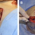
Surgical Technique for ADM Placement
- •
Immediately after completion of the mastectomy, a subpectoral pocket is developed.
- •
A thick piece of adequate-sized ADM is hydrated in normal saline.
- •
The breast mound pocket is measured and the appropriate tissue expander is selected.
- •
The ADM is then sutured to the remnant of the inframammary fold inferiorly and the lateral aspect of the chest wall (lateral mammary fold).
- •
The ADM is trimmed as it is inset to tailor the inferolateral sling to appropriate dimensions.
- •
The tissue expander is inserted in the submuscular/sub-ADM pocket and the superior border of the ADM sling is approximated to the inferior border of the pectoralis major muscle with 2-0 vicryl suture.
- •
Before placing the tissue expander, the air is evacuated from the device and 60 mL of injectable saline are added.
- •
After placement, an additional amount of injectable saline is added depending on the quality and amount of mastectomy flap skin.
- •
One or two closed suction drains are left in the mastectomy space.
- •
Any obviously devitalized skin is resected before closure.
- •
Patients are given prophylactic perioperative antibiotics, typically until drains are removed.
- •
Drains are removed when output is <30 cc/24 hours (usually 7-14 days post operatively).
- •
Expansion begins ∼14 days post operatively.
- •
Expansion continues every 1-4 weeks lateruntil complete.
Cost
A concern that many plastic surgeons have is the cost of incorporating ADM into breast reconstruction. Evidence suggests that the cost of a 1-stage reconstruction with ADM is similar to that of more traditional reconstructive techniques.
Janelle and colleagues compared the expected cost of immediate implant reconstruction using ADM with a 2-stage breast reconstruction without ADM. The study took into consideration complication rate, capsular contracture rate, operating room time, and the cost of ADM. Their analysis projected that a 1-stage reconstruction with ADM is about $500 less than a 2-stage reconstruction with a tissue expander without ADM. The study has limitations. The analytical model was based on estimated cost at a Canadian university-based medical center. Cost estimates may be different in other health care systems. Moreover, in the model presented, if a direct-to-implant case takes 30 minutes longer than immediate insertion of tissue expanders, the cost advantage of the single-stage procedure is negated.
For a more detailed review of costs related to ADMs, see the article by Macadam and Lennox elsewhere in this issue.
Related posts:
 Current State of the Art for Acellular Dermal Matrices in Breast Surgery
 Acellular Dermal Matrices in Breast Implant Surgery: Defining the Problem and Proof of Concept
 Pocket Reinforcement Using Acellular Dermal Matrices in Revisionary Breast Augmentation
 The Use of Human Acellular Dermal Matrices in Irradiated Breast Reconstruction
 Emerging Applications for Acellular Dermal Matrices in Mastopexy
Comparison of Different ADM Materials in Breast Surgery
Emerging Applications for Acellular Dermal Matrices in Mastopexy
Comparison of Different ADM Materials in Breast Surgery
Stay updated, free articles. Join our Telegram channel

Full access? Get Clinical Tree


