This chapter catalogues many examples of how the skin reacts to the external environment from which it protects the body. Exposure to physical factors such as heat, cold, moisture, ultraviolet light, radiation, mechanical trauma and imbedded foreign bodies can manifest as uniquely patterned skin findings.
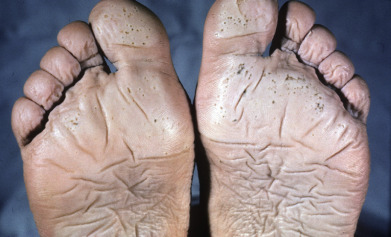
Of particular note are the many examples of photo-distributed eruptions, symmetrically involving the face, chest, dorsal hands and forearms, as this pattern can be an important physical exam clue to one of many specific photosensitive conditions. Those who spend significant amounts of time outdoors are particularly susceptible to acute and chronic sun damage and may also sustain temperature-related (cold or hot) injuries at acral sites.
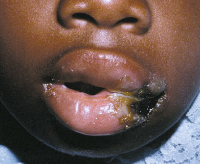
The distribution of mechanical injuries and foreign body reactions, however, is usually asymmetric. In this chapter, appreciate the geometric shapes and unnatural configurations that can be produced on the skin as a result of physical abuse, radiotherapy or phytophotodermatitis. The patient history, and at times tissue analysis, can help to confirm the diagnosis that was suspected after a thorough physical examination.
This portion of the atlas features examples of dermatoses and skin lesions resulting from physical factors.