Dermatoses of Pregnancy
Katrina Abuabara
Ellen K. Roh
Skin changes during pregnancy are common and range from physiologic changes to specific dermatoses of pregnancy. Some are well-defined conditions, such as pemphigoid gestationis, that can be diagnosed via direct immunofluorescence staining, while the etiology of many others remains poorly understood. There is a great deal of overlap in the terminology and diagnostic criteria for the dermatoses of pregnancy and even experienced dermatologists may have difficulty differentiating among some of the pruritic conditions. The information in this chapter is grouped into four categories: intrahepatic cholestasis of pregnancy, pemphigoid gestationis, polymorphic eruption of pregnancy (also commonly known as pruritic urticarial papules and plaques of pregnancy), and atopic eruptions of pregnancy, which includes eczema, prurigo of pregnancy, and pruritic folliculitis. Table 18-1 lists these categories with synonymous terms and associated conditions. Table 18-2 summarizes the three main conditions seen during pregnancy.
When evaluating a pregnant patient with a dermatologic complaint, physicians should inquire about gestational age, possibility of a twin pregnancy, parity, dermatologic changes in previous pregnancies, and a family history of pregnancy dermatoses. In addition to differentiating among dermatoses of pregnancy, the physician must rule out other common causes of cutaneous eruptions, such as allergic contact dermatitis, drug reactions, and insect bites. The physician should also be prepared to counsel the patient regarding potential effects to the fetus, cosmetic appearance, expected timing of resolution, and reoccurrence in subsequent pregnancies.
Physiologic Skin Changes During Pregnancy
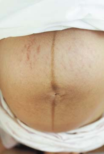
Hormonal and endocrine factors trigger a number of physiologic skin (Table 18-3), hair, connective tissue, and vascular changes during pregnancy. Hyperpigmentation affects up to 90% of pregnant women and includes darkening of the areolae, nipples, genital skin, axillae, and face (otherwise known as melasma or the “mask of pregnancy”). Melasma may also occur among women on oral contraceptives and can be exacerbated by sunlight. Patients should be advised to use sunscreen and to avoid excessive sun exposure. The linea alba becomes the linea nigra (Fig. 18-1) in the second trimester. Historically, it was assumed that darkening moles were also a physiologic, common change; however, it has recently been found that melanocytic nevi do not typically change during pregnancy. Therefore, any changing nevus in a pregnant patient should be closely observed and possibly biopsied.
Hirsutism, which is common on the face and occasionally on the extremities and back, affects many women during pregnancy. It typically resolves by the third trimester or in the postpartum period. Diffuse alopecia, or telogen effluvium, may begin a month or two postpartum and persist for up to a year. Striae distensae, “stretch marks,” appear as purple atrophic bands most commonly on the abdomen in the majority of women. Vascular changes such as spider telangiectasias; palmar erythema; nonpitting edema of the face, eyelids, and extremities; and hyperemia of the gums may also occur.
Hirsutism, which is common on the face and occasionally on the extremities and back, affects many women during pregnancy. It typically resolves by the third trimester or in the postpartum period. Diffuse alopecia, or telogen effluvium, may begin a month or two postpartum and persist for up to a year. Striae distensae, “stretch marks,” appear as purple atrophic bands most commonly on the abdomen in the majority of women. Vascular changes such as spider telangiectasias; palmar erythema; nonpitting edema of the face, eyelids, and extremities; and hyperemia of the gums may also occur.
Table 18-1 Dermatoses of Pregnancy and Common Synonyms | ||
|---|---|---|
|
Table 18-2 Differentiating the Dermatoses of Pregnancy | ||||||||||||||||||||||||||||
|---|---|---|---|---|---|---|---|---|---|---|---|---|---|---|---|---|---|---|---|---|---|---|---|---|---|---|---|---|
|
Table 18-3 Common Skin Changes in Pregnancy | |
|---|---|
|
Intrahepatic Cholestasis of Pregnancy (ICP)
A 25-year-old G1P1 white woman presents at 34 weeks of pregnancy with severe pruritus, especially of her hands and feet. She reports that she feels “exhausted,” though she thinks that it is because she has been unable to sleep the past few nights. She has also noticed that her urine is darker than usual. Her past medical history is notable for hepatitis C, treated with interferon. Physical examination reveals no primary lesions, only multiple excoriations.
Background/Epidemiology
Intrahepatic cholestasis of pregnancy (ICP) is characterized by a generalized pruritus and elevated bile acid levels consistent with cholestasis. It is unique in that there are no primary skin lesions associated with the condition, however, women may present with excoriations from scratching due to the intense pruritus. It is a rare condition; prevalence estimates range from 1 in 1,000 to 1 in 10,000 pregnancies, though rates are much higher in some geographic areas including Sweden and Chile.
Key Features
ICP presents as severe pruritus, particularly on hands and feet.
There are no primary skin lesions, only secondary change such as excoriations/crusts.
Other common features of presentation include dark urine color and light-colored feces.
Presents most commonly in the third trimester of pregnancy.
Treatment is with topical antipruritics for mild cases. Moderate or severe cases often improve with urso-deoxycholic acid therapy.
Pathogenesis
The etiology is heterogenous and thought to be caused by a combination of genetic, hormonal, and environmental factors. Multiple pregnancies and a history of liver damage are also thought to be risk factors.
Clinical Presentation
ICP typically presents during the third trimester of pregnancy as a generalized pruritus that may begin in the palms and soles and is usually more
severe at night. Patients can also have dark-colored urine, light-colored stool, and symptoms of fatigue, loss of appetite, and depression. Jaundice is present in about 20% of cases, and is associated with the development of cholesterol gallstones. In rare cases, ICP can cause steatorrhea, leading to decreased absorption of fat-soluble vitamins, vitamin K deficiency, and subsequent hemorrhage.
severe at night. Patients can also have dark-colored urine, light-colored stool, and symptoms of fatigue, loss of appetite, and depression. Jaundice is present in about 20% of cases, and is associated with the development of cholesterol gallstones. In rare cases, ICP can cause steatorrhea, leading to decreased absorption of fat-soluble vitamins, vitamin K deficiency, and subsequent hemorrhage.
ICP can be very uncomfortable because of severe pruritus, but poses few risks to the mother except in rare cases of steatorrhea and postpartum hemorrhage. It has been shown, however, to cause placental anoxia leading to fetal distress, meconium staining, preterm delivery, and even intrauterine fetal death.
Diagnosis
Liver function tests should be ordered for all pregnant women who experience pruritus. A rise in serum bile acid levels is the most sensitive indicator of ICP, and mild increases in alkaline phosphatase and transaminase levels occur in up to 60% of patients. ICP is a diagnosis of exclusion, therefore hepatitis panels should be sent to rule out a viral cause of liver disease. An abdominal ultrasound should be considered in patients with right upper quadrant symptoms to rule out cholelithiasis. Histopathology from skin biopsies are nonspecific and liver biopsy reveals nondiagnostic cholestatic changes.
Stay updated, free articles. Join our Telegram channel

Full access? Get Clinical Tree


