Abstract
The cutaneous deposition disorders are a heterogeneous group of conditions characterized by the presence of primarily endogenous substances within the dermis and/or the subcutis. These include gout, pseudogout, lipoid proteinosis, colloid milium, and mucopolysaccharidoses. Deposition disorders can be associated with localized or generalized cutaneous findings, and skin involvement can be the earliest sign of a systemic deposition disease. Histologic examination of cutaneous lesions, with the use of special stains, is a very helpful diagnostic tool. In addition, specific enzymatic assays or genetic mutational analysis can then be performed to establish the precise diagnosis.
Keywords
gout, pseudogout, lipoid proteinosis, colloid milium, mucopolysaccharidoses, cutaneous deposition disease, urate crystals, birefringence, tophi
Introduction
The cutaneous deposition disorders are a heterogeneous group of conditions characterized by the presence of primarily endogenous substances within the dermis or the subcutis. Deposition disorders can be associated with localized or generalized cutaneous findings, and skin involvement is sometimes the earliest sign of a deposition disease. Histologic examination of cutaneous lesions, with the use of special stains including immunohistochemistry, is a very helpful diagnostic tool ( Table 48.1 ). In addition, specific enzymatic assays or genetic mutational analysis can then be performed to establish the precise diagnosis.
| DISORDERS WITH DERMAL DEPOSITS – HISTOLOGIC FEATURES | |||
|---|---|---|---|
| Disease | Microscopic findings | Histochemical stains | Special processing |
| Gout | Dermal deposits of amorphous material with needle-like clefts surrounded by giant cells Polarizing light/fresh preparation – fine, needle-like crystals; negative birefringence | 20% silver nitrate solution: urate crystals appear black and the surrounding tissue yellow De Galantha stain: crystals brown–black | Best fixative to preserve crystals: ethanol-based (Carnoy’s fluid) |
| Lipoid proteinosis | Masses of amorphous or laminated hyalin-like material Also surrounds capillaries in the papillary dermis and appendages | Contains type IV collagen and laminin PAS-positive and diastase-resistant ⊕ Alcian blue, hyaluronidase-sensitive Sudan III (+/−) Congo red (usually −) | Direct immunofluorescence with anti-ECM1 antibodies to assess protein expression |
| Colloid milium (adult form) | Nodules of amorphous eosinophilic material Artifactual clefts Solar elastosis | Stains similarly to amyloid (but not all cases) ⊕ Congo red ⊕ Thioflavin T ⊕ Crystal violet | |
| Mucopolysaccharidoses | Granules within fibroblasts Large vacuolated cells in sweat glands and the external root sheath of hair follicles (gargoyle cells) | Granules within fibroblasts: ⊕ Metachromasia with toluidine blue and Giemsa ⊕ Alcian blue ⊕ Colloidal iron (see Table 46.1 ) | Fixation in absolute alcohol better than in formalin (consider when clinical diagnosis not supported by histologic findings) |
| Mucinoses, e.g. papular mucinosis, scleredema, pretibial myxedema ( Ch. 46 ) | Mucin between collagen and variable fibroblast proliferation | ⊕ Metachromasia with toluidine blue ⊕ Alcian blue ⊕ Colloidal iron (see Table 46.1 ) | Fixation in absolute alcohol better than in formalin |
| Amyloidosis ( Ch. 47 ) | Fractured, amorphous, faintly eosinophilic material in dermis | ⊕ Congo red ⊕ Thioflavin T ⊕ Crystal violet | In glutaraldehyde for electron microscopy |
| Ochronosis ( Ch. 67 ) | Sausage- or banana-shaped brown/yellow/orange structures | Black staining with methylene blue or cresyl violet | |
| Calcinosis cutis ( Ch. 50 ) | Irregular masses of deeply basophilic material Variable granulomatous inflammation | ⊕ von Kossa ⊕ Alizarin red (more specific) | May fracture microtome blade May need decalcification solution first |
| Porphyrias, in particular EPP ( Ch. 49 ) | Perivascular rim of delicate eosinophilic fibrillar material In chronic lesions, also dense homogeneous deposits | PAS-positive and diastase-resistant Immunohistochemistry for BMZ components (type IV collagen) | |
This chapter will focus on gout, pseudogout, lipoid proteinosis, colloid milium, and the mucopolysaccharidoses. Additional deposition disorders such as papular mucinosis, amyloidosis, porphyria, and calcinosis cutis are reviewed in Chapter 46 , Chapter 47 , Chapter 49 , Chapter 50 .
Gout
▪ Urate crystal arthropathy ▪ Podagra ▪ Urate deposition disease
- ▪
Gout is a metabolic disease in which the underlying abnormality is hyperuricemia
- ▪
Deposits of needle-like crystals of monosodium urate are found in several sites, most commonly the skin (as tophi) and the joints
- ▪
In fresh preparations of synovial fluid and expressed material from tophi, the crystals exhibit negative birefringence when examined by polarized light
- ▪
In routinely fixed histologic sections, amorphous deposits that contain needle-like clefts are seen in the dermis and subcutis
Introduction
Gout is a metabolic disease in which needle-like crystals of monosodium urate (the ionized form of uric acid) from supersaturated fluids are deposited in tissue. Clinical manifestations include gouty arthritis, accumulation of crystals in connective tissue (tophi), uric acid nephrolithiasis, and renal impairment .
Epidemiology
Gout is the most common form of crystal-induced arthritis, and its prevalence seems to have increased over the past few decades. Men 40–50 years of age are most commonly affected. The male-to-female ratio is 9 : 1, and most women with gout are postmenopausal. Gout is found in up to 4% of adults and ~20% of patients have a family history of gout .
Pathogenesis
Gout is a consequence of deposits of urate crystals that have precipitated from supersaturated body fluids. Uric acid is the end product of the catabolism of purines. When the body increases its production of uric acid or if the kidneys do not excrete sufficient amounts, the result is hyperuricemia ( Table 48.2 ).
| CLASSIFICATION OF HYPERURICEMIA | |
|---|---|
| Overproduction of uric acid |
|
| Decreased excretion of uric acid |
|
Urate crystals can activate the NLRP3 (cryopyrin) inflammasome (see Fig. 4.2 ) and stimulate the production of the proinflammatory cytokine interleukin-1β by monocytes and macrophages . The subsequent migration and activation of neutrophils as well as complement activation initiate a vicious cycle. The ingestion of crystals by neutrophils triggers cell damage and leakage of lysosomes, which then result in further inflammation and tissue damage.
Clinical Features
The typical patient with gout is a middle-aged or older man, who may have a family history of the disease. Risk factors include obesity, excessive alcohol intake, renal insufficiency and certain medications, most commonly diuretics. The natural history of gout involves four clinical stages : (1) asymptomatic hyperuricemia; (2) acute gouty arthritis; (3) intercritical gout (the intervals between attacks, which progressively become shorter in duration); and (4) chronic tophaceous gout.
Acute gouty arthritis
The most common symptom of gout is the development of severe joint pain over a period of 6–12 hours, accompanied by tenderness, erythema, warmth, and swelling. Initial attacks are often monoarticular, but in up to 40% of patients they may be polyarticular . In 75% of patients, the first metatarsophalangeal joint is involved (referred to as podagra); additional sites include the knee, ankle and foot, and, less commonly, the hand, wrist and elbow. Fever and systemic symptoms may accompany an acute attack of gout. As the swelling subsides, the skin acquires a violaceous hue often followed by desquamation.
Chronic tophaceous gout
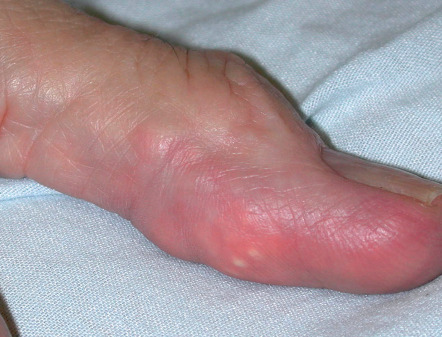
Cutaneous deposits of monosodium urate, known as “tophi”, usually appear an average of 10 years after the onset of gout. These tophi present as firm dermal or subcutaneous papules and nodules or as a fusiform swelling. Their contours may be smooth or multilobulated ( Fig. 48.1 ) and they vary from skin-colored to white–yellow to red ( Fig. 48.2 ). The surface may be ulcerated and there may be associated drainage of material, which varies from clear fluid with white flecks to a thick chalky material.
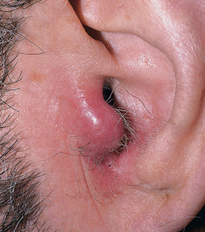
The most common locations for tophi are the skin overlying joints and the helix of the ear. Unusual sites include the eyes, nose, larynx, breast, and heart valves. However, tophi develop in fewer than 10% of patients with gout. Depending upon the size and location of the tophi, complete, partial or minimal resolution can occur when the serum uric acid level is normalized .
Elevated levels of uric acid in the urine are associated with uric acid nephrolithiasis and may cause acute renal failure when uric acid precipitates in the renal tubules and collecting ducts. The most common setting for the latter is the tumor lysis syndrome in which chemotherapy is given for a rapidly dividing chemosensitive malignancy, e.g. leukemias, lymphomas.
The initial diagnosis of gout is usually made in the setting of acute arthritis, based upon the presence of urate crystals in an aspirate of joint fluid. Hyperuricemia is usually present, but alone it is insufficient for establishing the definitive diagnosis of gout. Additional laboratory abnormalities during an acute attack include an elevated white blood cell count (in the joint fluid and blood) and an elevated ESR.
When examined by polarized light microscopy, the needle-shaped urate crystals change color from yellow to blue based upon their alignment relative to the axis of the “red plate” compensator, i.e. they exhibit negative birefringence. In patients with recurrent attacks, radiographs of involved joints often show “punched-out” erosions with sclerotic “overhanging” margins, but no osteophytes. A 24-hour urine collection for uric acid allows the identification of patients at risk for nephrolithiasis (see Table 48.2 ).
Pathology
In gouty tophi, the key histologic feature is the presence of deposits of amorphous material within the dermis and subcutis. These deposits contain needle-like clefts (which represent dissolved urate crystals) surrounded by an infiltrate composed of histiocytes, multinucleated giant cells, and lymphocytes ( Fig. 48.3 ). Secondary calcification or even ossification can occasionally be seen.
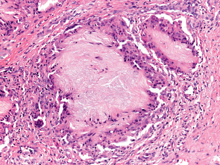
In order to preserve the crystals, the biopsy specimen needs to be placed in an ethanol-based fixative, such as Carnoy’s fluid. Then, under regular or polarized light, brightly refractile brown sheaths of fine needle-like crystals can be seen. Upon staining with 20% silver nitrate solution, the crystals appear black and the surrounding tissue yellow, whereas with the De Galantha stain, the crystals appear brown to black .
Differential Diagnosis
The differential diagnosis of acute gouty arthritis includes pseudogout (see below), osteoarthritis, psoriatic arthritis, reactive arthritis (previously referred to as Reiter disease), and septic arthritis. Given its destructive nature, septic arthritis needs to be considered even in patients in whom the diagnosis of gout has been previously established. The presence of needle-shaped crystals with negative birefringence in an aspirate of joint fluid is diagnostic of gout, while Gram stain and culture exclude septic arthritis. Occasionally, patients are misdiagnosed as having cellulitis.
The differential diagnosis of cutaneous gouty tophi includes xanthomas, rheumatoid nodules, and calcinosis cutis. Examination of expressed fluid or chalky material by polarized light can provide a “bedside” diagnosis. By ultrasonography, central clear spaces are seen within tophi whereas rheumatoid nodules contain central echodense areas .
Treatment
For acute gout, short-acting oral nonsteroidal anti-inflammatory drugs ( NSAIDs ) are taken within 24 hours of onset at the highest safe dosage as long as symptoms persist, e.g. indomethacin 50 mg three times daily for several days. Contraindications include peptic ulceration, renal insufficiency, and anticoagulation .
Colchicine , a derivative of the autumn crocus, has been used to treat gout for centuries. This drug relieves pain and swelling and can help to prevent future attacks. Colchicine inhibits the transport of phagocytized urate crystals to lysosomes within neutrophils via its binding to microtubules. Colchicine also interferes with leukocyte migration, chemotaxis, and adhesion. For patients with normal renal function who are not receiving strong CYP3A4 inhibitors, the Food and Drug Administration (FDA)-recommended dosage for acute gout flares is 1.2 mg orally followed by 0.6 mg 1 hour later. Low-dose NSAIDs and colchicine (0.6 mg twice daily) are both utilized for prophylaxis. Side effects of colchicine are outlined in Table 130.10 . Intra-articular or systemic corticosteroids may be used for a period of 1–3 weeks if NSAIDs and colchicine are ineffective or contraindicated .
The xanthine oxidase inhibitors allopurinol and febuxostat block uric acid production and are used primarily for chronic gout; in the setting of acute gout, effective anti-inflammatory therapy is first established . Side effects of allopurinol include diarrhea, thrombocytopenia, hepatitis, and skin eruptions that can range from morbilliform to erythroderma to Stevens–Johnson syndrome/toxic epidermal necrolysis (SJS/TEN). HLA-B*58:01-positive individuals are at markedly increased risk of developing severe cutaneous adverse reactions, including both SJS/TEN and DRESS. Of note, this allele is most common in those of Han Chinese, Korean or Thai descent. Major side effects of febuxostat are liver enzyme elevations and cardiovascular thromboembolic events . Drug–drug interactions, e.g. with azathioprine, can occur with both of these medications (see Fig. 130.4 ). Uricosuric agents such as oral probenecid can be added or used as second-line therapy for patients with normal renal function and no renal stones. Overall, maintenance of a serum uric acid level of ≤6 mg/dl is recommended. A newer agent, lesurinad, is a selective inhibitor of reabsorption of uric acid.
Some patients may benefit from a reduction in purine-rich foods (e.g. organ meats [liver, heart], fish), alcohol consumption, and body weight . In the setting of potential tumor lysis syndrome, intravenous rasburicase can be administered, which rapidly converts uric acid to the more soluble allantoin. Bimonthly intravenous pegloticase , which has the same mechanism of action, is approved for patients with chronic gout refractory to conventional therapy, and improvement in tophi has been observed. IL-1 blocking agents (e.g. anakinra, canakinumab ) have been used for recurrent acute gout unresponsive to other therapies, but are not currently FDA-approved for this indication (see Fig. 45.13 ).
Related posts:


Stay updated, free articles. Join our Telegram channel

Full access? Get Clinical Tree


