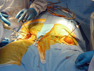
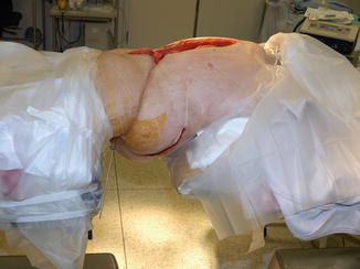
Fig. 19.1
Clinical photograph of patient with myelomeningocele, two-level infected pseudarthrosis of the lumbar spine, and morbid obesity. Fixation consists of pelvic wire and half-pin fixation, thoracic spinal fixation with pedicle half-pins, and a halo ring
19.5 Imaging
Plain sitting AP and lateral radiographs of the spine are necessary to document the severity, nature, and location of spinal deformity.
Axial CT of the intended area of fixation, specifically the lumbosacral spine and pelvis, and the thoracic spine in the intended area of thoracic pedicle fixation is essential (Fig. 19.2a, b). If necessary for the specific case, CT of the mid-lumbar spine with or without reconstructions may be helpful (Fig. 19.3).
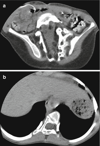
Fig. 19.2
(a, b) Axial CT of the intended areas of fixation is necessary to identify bone stock and shape in preparation for fixation. (a) Axial CT of the pelvis, for anticipated posterior-to-anterior open-wire fixation. (b) Axial CT of the thoracic spine at the level of intended posterior pedicle half-pin fixation
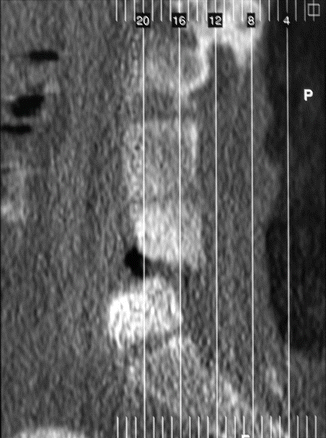
Fig. 19.3
Sagittal CT reconstruction of the lumbar spine identifying two-level lumbar pseudarthrosis and nature of residual sagittal deformity. Same patient as Fig. 19.1
In patients with myelomeningocele and ventriculoperitoneal (VP) shunts, it is important to know the functional status of the shunt at the time of surgery. Patients who are shunt-dependent and being considered for residual spinal cord resection as part of the indicated spinal release must have a functioning shunt prior to that procedure.
19.6 Special Equipment Required
Custom body rings or equivalent
Extra length and diameter wires for large patients
Special wire fixation bolts for larger-diameter wires
Operating table setup as described below
Special bed preparations as described below
Dedicated staff to provide the full-time nursing care necessary during the prolonged hospitalization required to achieve a successful outcome in these extraordinarily challenging cases
19.7 Frame Construction
We now use a custom ring or similar to fashion circular fixation of the pelvis, which will serve as the foundation to the entire construct (Fig. 19.4).
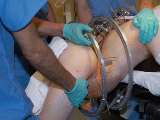
Fig. 19.4
A 360 mm custom-made aluminum ring, which we have used as both pelvic and “float” rings in our spinopelvic apparatus
The upper segment consists of a series of arches to which the thoracic pedicle half-pins are fixed as a block (Fig. 19.5).
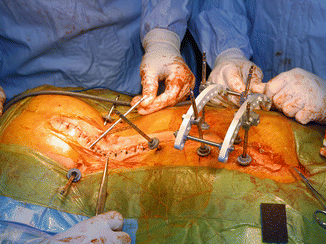
Fig. 19.5
Thoracic pedicle half-pins secured to an upper femoral arch
The pelvic ring and thoracic pedicle block are attached to each other in a manner required for the specific case: fixed in an acceptable position for patients with mobile-infected pseudarthrosis of the spine and with parallel hinges located under fluoroscopic control and “motors” used in either distraction or compress to effect gradual correction of hyperlordotic or kyphotic deformities (Fig. 19.6).
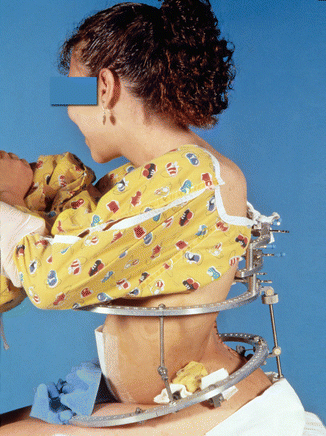
Fig. 19.6
Clinical photograph of a patient with hyperlordosis of the spine undergoing gradual correction of residual deformity. The upper segment consists of thoracic pedicle half-pins secured to arches, the lower segment of pelvic wires and half-pins secured to a full-body ring, and there is a “float” body ring between the two segments to facilitate hinge and motor connection between the segments
19.8 Intraoperative Patient Positioning
Any initial indicated procedure (such as anterior and/or posterior spinal release or wound debridement) is carried out in a routine fashion per the surgeon’s preference.
For spinopelvic fixation, we position our patients prone between two operating tables, one to support the thorax and the other to support the legs from the lower pelvic area. The operating tables should be radiolucent to allow fluoroscopic imaging to aid in pedicle half-pin insertion. Split draping of the patient’s upper and lower body segments will then allow circumferential access to the patient’s pelvis and abdomen (Fig. 19.7).
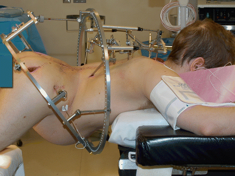
Fig. 19.7
Intraoperative patient positioning for spinopelvic fixation. Two operating tables are used, with the patient’s thorax supported on one and the legs on a second table, allowing circumferential access to the pelvis and abdomen. Tabletops should be radiolucent to allow fluoroscopic imaging during fixation element insertion
When we use anteriorly placed iliac half-pins to supplement pelvic fixation, we insert them first through anterior exposure of the pelvis (Fig. 19.8) and then turn the patient prone with re-prepping and draping as described above (Fig. 19.9).
