Case 19 Prominent Ear Deformity
19.1 Description
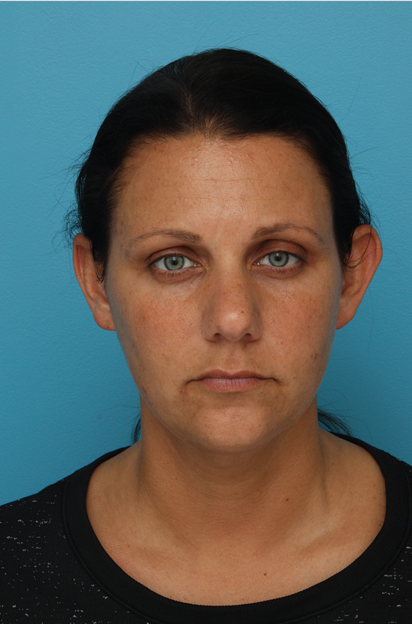
Adult patient with evidence of side asymmetry with the left ear more prominent than right
The antihelical fold (scaphoconchal angle) appears obtuse with increased projection of the scapha and helix
19.2 Work-Up
19.2.1 History
Patient’s age
Patient’s goals and expectations
History of prior surgery to ears
History of prior ear trauma
History of poor wound healing (e.g., prior keloids)
19.2.2 Physical Examination
Presence/absence of preoperative asymmetry
Overall size and shape of ear
Helix–mastoid angle
Helix–mastoid distance (upper, middle, and lower thirds of the ear)
Cartilage consistency
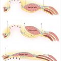
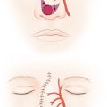
Characteristics to be evaluated (Fig. 19-1)
Upper third of the ear
Underdeveloped/deficient antihelical fold
Obtuse scaphoconchal angle (>90 degrees)
Middle third of the ear
Conchal hypertrophy (concha cavum >1.5 cm deep)
Obtuse conchal–mastoid angle (>25 degrees)
Lower third of the ear
Related posts:
Stay updated, free articles. Join our Telegram channel

Full access? Get Clinical Tree


