Abstract
Cutaneous deposition of calcium has a range of manifestations, from mild to fatal, and a number of etiologies. It is categorized as dystrophic if calcium deposits in pre-existing damaged skin, metastatic if it results from a calcium and/or phosphate imbalance, mixed if both dystrophic and metastatic factors converge, iatrogenic if due to medical interventions, and idiopathic if no cause can be determined. When calcium combines with phosphorus and deposits as hydroxyapatite crystals in a proteinaceous matrix, osteoma cutis develops. Osteoma cutis can arise in association with chronic acne and as a manifestation of several genetic disorders. The treatment of these conditions can be challenging, therefore early identification and intervention are critical.
Keywords
calcinosis cutis, osteoma cutis, metastatic calcification, dystrophic calcification, mixed calcification, idiopathic calcification, iatrogenic calcification, calciphylaxis
- ▪
Calcium regulates several key physiologic events in the skin, including epidermal proliferation, differentiation, and cell–cell adhesion
- ▪
Disruption of normal calcium regulatory pathways can lead to calcification and/or ossification of the skin
- ▪
Five major types of cutaneous calcification are seen: dystrophic , in which pre-existing damage to the skin results in calcium deposits; metastatic , where systemic metabolic disorders lead to calcium deposits in the skin and often other tissues; iatrogenic , as a result of medical therapy or testing; idiopathic , in which no cause can be identified; and mixed , a combination of metastatic and dystrophic
- ▪
In cutaneous ossification, there is deposition of calcium and phosphorus as hydroxyapatite crystals within a protein matrix; it can occur in conjunction with calcification or primarily in a number of genetic syndromes
- ▪
For correct classification of calcifying disorders, an evaluation of calcium-phosphate metabolism and an examination for associated systemic abnormalities are required
- ▪
Treatment of calcifying disorders is difficult. Correction of any associated disorder, such as hyperparathyroidism, is essential to prevent further calcification. Efficacy of agents that modify calcium metabolism is based primarily upon case series. Surgical excision may be useful in circumscribed disease that interferes with function
Introduction
Calcium plays a vital role in regulating key physiologic events in many tissues, including the skin. In the epidermis, calcium shares in the control of major functions, including proliferation, differentiation, and cell–cell adhesion. Control of intra- and extracellular calcium concentrations and maintenance of gradients are essential for its regulatory role. When factors that regulate calcium in the skin are disrupted, by either local or systemic events, cutaneous calcification or ossification as well as acantholysis and dyskeratosis can develop.
Calcification arises from the deposition of amorphous, insoluble calcium salts, while ossification results from deposition of calcium and phosphorus in a proteinaceous matrix as hydroxyapatite crystals. Ossification may follow pre-existing calcification in the skin, but in most disorders one form of calcium deposition predominates. Each of the calcifying and ossifying disorders of the skin is rare, but as an aggregate finding calcium deposition within the skin is not uncommon.
Cutaneous Calcification
Calcifying disorders of the skin are generally divided into five broad categories: dystrophic, metastatic, idiopathic, iatrogenic, and mixed calcification . Dystrophic calcification occurs in the setting of localized tissue damage, without an underlying metabolic abnormality in calcium regulation. Theoretically, dystrophic calcification occurs because the underlying disease process damages cell membranes, allowing calcium influx and subsequent intracellular crystallization. Alternatively, the acidity that accompanies cell damage may disrupt normal processes that inhibit calcification. In contrast, metastatic calcification occurs in normal tissue when there is dysfunction of calcium regulatory systems. When no known local or systemic factors can be identified, the calcification is categorized as idiopathic , and that related to medical therapy or testing is iatrogenic . Mixed calcification arises when metastatic calcification due to an underlying metabolic abnormality acts as a trigger for dystrophic calcification.
Dystrophic Calcification
Autoimmune Connective Tissue Disease
Dystrophic calcification may be seen in any of the autoimmune connective tissue diseases (AI-CTDs), but it is most common in childhood dermatomyositis and in the CREST form of systemic sclerosis. As a comparison, cutaneous calcification develops in 10–20% of patients with adult dermatomyositis, whereas it occurs in ~50–70% of children with the disease (see Fig. 42.8 ) . The presence of dermatomyositis-associated calcinosis correlates with longer disease duration, fingertip ulceration, and anti-NXP-2 autoantibodies, while a negative correlation exists with anti-transcriptional intermediary factor 1-γ antibodies . Small deposits may occur in the skin or larger firm masses may begin in the muscle groups most severely affected by the disease. The most frequently involved sites are the elbows, knees, buttocks, and shoulders. Extrusion of calcium through the skin causes significant morbidity with pain and secondary infection.
In calcinosis universalis, the most severe form of dystrophic calcification, there is diffuse involvement with sheet-like masses of calcium. Calcification occurs along fascial planes, often leading to severe functional impairment. Aggressive treatment of dermatomyositis with immunosuppressive agents and/or IVIg may be warranted to try to limit this serious complication (see Ch. 42 ). However, once calcification has developed, the effectiveness of these therapies can be quite variable.
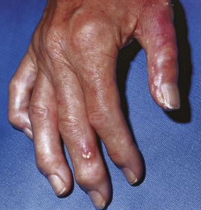
The CREST ( c alcification, R aynaud phenomenon, e sophageal dysmotility, s clerodactyly, and t elangiectasia) variant of systemic sclerosis is the other AI-CTD in which cutaneous calcification is frequently seen. Calcification is usually less severe than in dermatomyositis and is usually restricted to the hands and upper extremities ( Fig. 50.1 ); it is also seen over bony prominences and tendons. Extrusion of white chalky material is sometimes followed by localized ulceration, often at sites of trauma such as the digits. Calcinosis universalis can occur in systemic sclerosis, but much less frequently than in dermatomyositis.
Dystrophic calcification has occasionally been reported in chronic cutaneous, acute systemic, and subacute cutaneous lupus erythematosus as well as generalized morphea ( Fig. 50.2 ) . Calcification may also occur within lesions of lupus panniculitis. In systemic lupus erythematosus, calcification is most often an asymptomatic radiologic finding.
Treatment of dystrophic calcification
Therapies for dystrophic calcification include a low calcium and phosphate diet, aluminum hydroxide, and bisphosphonates, although no controlled trials have convincingly shown clinical improvement. In case reports and small series of patients, colchicine, probenecid, and sodium thiosulfate have demonstrated some efficacy. For small deposits (<3mm), application of topical sodium thiosulfate (12.5 mg/ml compounded 1:1 with petrolatum applied daily) can lead to resolution, while larger lesions may respond to intradermal injections (12.5 mg/ml, 0.1–1 ml every 3 to 6 weeks). Long-term treatment with diltiazem was reported to decrease the size of calcium deposits, presumably through its effect on calcium transport into cells . However, months to years of treatment may be required for extensive calcification. Surgical excision is appropriate in selected patients with localized masses that are painful or interfere with function, but recurrence can occur.
Panniculitis
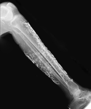
Any lobular panniculitis may develop focal dystrophic calcification, including pancreatic panniculitis, lupus profundus, and subcutaneous fat necrosis of the newborn (see Ch. 100 ). In the latter, the calcium deposits are small and are most easily noted radiologically; occasionally, more widespread deposits may be seen .
Genetic Disorders
A number of genetic disorders may be accompanied by cutaneous calcification, including pseudoxanthoma elasticum (PXE; see Ch. 97 ). The coexistence of typical elastic fiber calcification along with small discrete foci of cutaneous calcification or ossification has been reported . PXE results from inactivating mutations in ABCC6 which encodes a membrane transporter system . ABCC6 directly mediates the sinusoidal release of ATP from the liver, which is then converted to AMP and inorganic pyrophosphate (PPi) within the liver vasculature . PPi normally prevents the matrix calcification that is thought to create the findings in PXE . In addition, serum levels of fetuin-A, another major anti-mineralization protein, are significantly reduced in PXE patients . These factors, combined with impaired carboxylation of the inhibitor matrix Gla protein, may allow for increased responsiveness to pro-calcifying stimuli and unchecked calcification of elastic fibers in affected tissues .
Ehlers–Danlos syndrome encompasses multiple disorders in which genetic mutations lead to abnormal collagen synthesis, metabolism, or function (see Ch. 95 ). Patients develop hard subcutaneous nodules known as spheroids or spherules over bony prominences, which are thought to represent herniated fat lobules that have become calcified (see Ch. 97 ). It is also possible that minor trauma results in scar tissue which then becomes a focus for dystrophic calcification . Of note, similar calcifications can occur within the breast in these patients and their detection by mammography may lead to false-positive radiographic findings for breast carcinoma.
Dystrophic calcification may also be seen in patients with porphyria cutanea tarda (PCT; see Ch. 49 ). It occurs most often in patients with longstanding disease who develop sclerodermoid changes and secondary calcification within these areas . As with the other lesions of PCT, calcification appears most often in the head and neck region or on the dorsal surface of the hands. Ulceration has also been reported, although rarely, in association with these lesions.
Other rare genetic syndromes in which cutaneous calcification may be seen include Werner syndrome and Rothmund–Thomson syndrome (see Ch. 63 ). Cerebral amyloid angiopathy , an autosomal dominant condition due to mutations in the gene encoding the amyloid precursor protein, can present with dementia, patchy leukoencephalopathy, hemorrhagic stroke, external carotid artery dysplasia, and occipital calcifications . Microcalcification of the dermal blood vessels may also be seen, and while the latter is asymptomatic, dermatologists might be called upon to biopsy the skin as a diagnostic measure.
Infections
Infections, particularly parasitic, can lead to dystrophic calcification. Calcified cysts form around larvae or worms, including Onchocerca volvulus and the tapeworm Taenia solium . Intrauterine herpes simplex viral infections may cause annular plaques of calcinosis cutis in newborns.
Neoplasms
Incidental calcification has been noted in a large number of benign and malignant tumors of the skin. Up to 75% of pilomatricomas develop calcification and in 15–20% there is evidence of ossification. A chalky, calcium-containing material may drain from the surface of these tumors. Activating mutations in the β-catenin gene have been demonstrated in sporadic pilomatricomas . Other calcifying adnexal tumors or cysts include basal cell carcinomas, pilar cysts, epidermoid inclusion cysts, and chondroid syringomas . Rarely, melanocytic nevi, atypical fibroxanthomas, pyogenic granulomas, trichoepitheliomas, and seborrheic keratoses have been reported to calcify.
Other
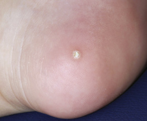
Trauma, “heel sticks” (for obtaining blood in neonates; Fig. 50.3 ), subcutaneous or intramuscular injections sites, surgical and burn scars, and keloids have also been reported as triggers of dystrophic calcification.
Metastatic Calcification
Renal Disease
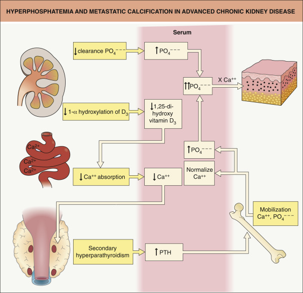
The most common cause of metastatic calcification is advanced chronic kidney disease ( Fig. 50.4 ). In patients with renal failure, there is an impaired ability to clear phosphate and impaired activation of vitamin D3, as 1-α hydroxylation occurs in the kidney. Impaired production of 1,25-dihydroxyvitamin D 3 leads to decreased absorption of calcium from the intestine and hypocalcemia. Hypocalcemia, in turn, induces increased secretion of parathyroid hormone (PTH) and subsequent mobilization of calcium and phosphate. The serum calcium concentration is normalized, but significant hyperphosphatemia may develop, and, if the solubility product of calcium × phosphate is exceeded, metastatic calcification can result.
Benign nodular calcification usually develops in the setting of prolonged secondary hyperparathyroidism due to advanced chronic kidney disease. Clinically, there are large deposits of calcium in the skin and subcutaneous tissue, often in periarticular sites. The number and size correlate with the severity of the hyperphosphatemia. The lesions are usually asymptomatic except for pain from pressure placed on surrounding structures. Normalization of serum calcium and phosphate levels may result in resorption of the lesions; however, if larger deposits interfere with function, surgical removal is recommended.
In renal transplant recipients, deposition of calcium within the skin has been reported following subcutaneous administration of low-molecular-weight heparin (nadroparin). Nodules with secondary ulceration developed at the sites of the heparin injections, but the process was self-limited and resolved following discontinuation of the nadroparin. The calcium content of the heparin in combination with hyperphosphatemia due to renal dysfunction was hypothesized as the underlying pathogenic mechanism .
Milk–Alkali Syndrome
Ingestion of excessive amounts of calcium-containing foods or antacids can lead to hypercalcemia and the milk–alkali syndrome. Patients with milk–alkali syndrome have acute manifestations including nephrocalcinosis, irreversible renal failure, and diffuse subcutaneous calcification.
Hypervitaminosis D
Chronic ingestion of supraphysiologic doses of vitamin D can lead to hypercalcemia and hypercalciuria. Clinical signs include weakness and lethargy, nausea, headache, and polyuria. Nephrolithiasis and calcinosis cutis may occur. Hypercalcemia is also seen in patients with sarcoidosis secondary to increased calcium absorption due to 1,25-dihydroxyvitamin D production by the granulomas. For other causes of metastatic cutaneous calcification, see Table 50.1 .
| DISORDERS OF CUTANEOUS CALCIFICATION |
| Dystrophic |
|
| Metastatic |
|
| Idiopathic |
|
| Iatrogenic |
|
* Can occasionally occur in the setting of severe primary hyperparathyroidism and, less often, in the absence of a clearly identifiable trigger.
Mixed Calcification
In mixed calcification, there is a combination of metastatic and dystrophic calcification, which is initiated by calcium dysregulation and then propagated by trauma.
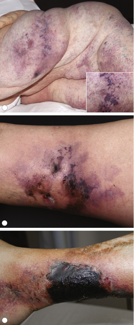
Calciphylaxis
Calciphylaxis is characterized by intimal fibrosis and medial vascular calcification (that can become transmural) as well as transdifferentiation of vascular smooth muscle cells into osteoblast-like cells; these changes plus thrombosis lead to ischemic necrosis of the skin and soft tissues . For vascular calcification to occur, an important step is the conversion of vascular smooth muscle cells into osteoblast-like cells. Phosphates, inflammatory mediators in the vessel wall, and bone morphogenic protein (BMP)-2 have all been proposed as potential stimuli for this conversion. In addition, BMP-7, matrix Gla protein, fetuin-A, osteoprotegerin, and phosphatonins are possible positive or negative regulators of this process .
Early lesions usually present as poorly demarcated, often symmetric, very painful patches of erythema or retiform purpura; they favor areas with abundant adipose tissue or sites of trauma ( Fig. 50.5A,B ). Bullae or a dusky gray color may then develop, signifying imminent tissue necrosis and the appearance of ulcerations with black, leathery eschars ( Fig. 50.5C ). Often, there is surrounding subcutaneous induration that extends beyond the margins of the visibly active lesions and this can be helpful in distinguishing calciphylaxis from other forms of retiform purpura. Death may occur from secondary infection and sepsis or internal organ involvement. One-year mortality rates range from 45% to 80%, with proximal lesion location and ulceration associated with a higher risk of death .