Abstract
For over thirty years, botulinum neurotoxins (BoNTs) have been used for therapeutic and cosmetic applications. Of the five formulations of BoNT type A (BoNT-A) available worldwide, three have been approved by the U.S. Food and Drug Administration: onabotulinumtoxinA, abobotulinumtoxinA, and incobotulinumtoxinA. One formulation of BoNT type B (rimabotulinumtoxinB) is also available. BoNT-A has been studied extensively for the treatment of rhytides of the face and neck. For improved aesthetic outcomes, it may be combined with other rejuvenation procedures such as soft-tissue augmentation or laser and other light-based therapies. Beyond the cosmetic field, BoNTs have been utilized for a number of therapeutic applications, including hyperhidrosis, migraine and Raynaud phenomenon, with great success. Side effects are typically mild and transient, especially when BoNT is used in small doses for cosmetic applications. Although more serious complications can occur, they are largely due to improper injection techniques or over-enthusiastic doses. Overall, BoNT carries an impressive record of safety, and its clinical uses continue to evolve.
Keywords
neuromodulators, botulinumtoxinA, onabotulinumtoxinA, incobotulinumtoxinA, abobotulinumtoxinA, rimabotulinumtoxinB, Botox ® , Xeomin ® , Dysport ® , rhytides, hyperhidrosis
- ▪
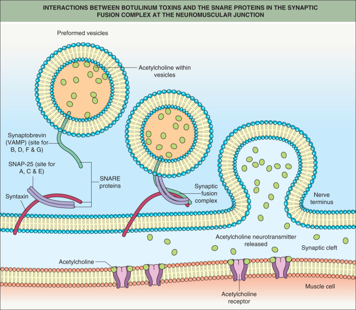
Botulinum toxin (BoNT) causes chemodenervation of muscles by blocking acetylcholine release
- ▪
BoNT is also capable of inhibiting the release of other neurotransmitters, such as substance P, noradrenaline, calcitonin gene-related peptide, and glutamate from a variety of synaptosomal and neuronal systems
- ▪
Injections of BoNT type A (BoNT-A) that weaken or relax muscles can smooth hyperfunctional lines and change the contour of the face and neck
- ▪
Adjunctive use of BoNT-A with soft-tissue augmentation or laser resurfacing is beneficial, particularly in active regions such as the perioral and periocular areas
- ▪
The cosmetic effects of BoNT-A typically last months and may increase with subsequent injections
- ▪
In addition to its effect on musculature, BoNT-A successfully denervates sweat glands, serving as an effective alternative treatment for hyperhidrosis, and it can also improve the pain, swelling, and discoloration associated with Raynaud phenomenon
- ▪
Complications (e.g. eyelid ptosis) are generally caused by diffusion of the toxin to non-targeted muscles and can be minimized by using concentrated doses and careful technique
Introduction
Botulinum toxin (BoNT) injections are commonly used to improve facial aesthetics by smoothing hyperdynamic rhytides of the face and neck. In recent years, the use of BoNT has evolved to reflect a more three-dimensional approach to facial rejuvenation, often in combination with soft-tissue augmentation or other restorative procedures. Moreover, a better understanding of its mechanism of action has led to innovative indications across a number of therapeutic fields.
Properties of Botulinum Toxins
Various strains of the bacterium Clostridium botulinum produce seven distinct serotypes of BoNT that affect neural function: A, B, C1, D, E, F and G . Although all serotypes produce chemodenervation and atrophy of skeletal muscles by blocking acetylcholine release from motor neurons at the neuromuscular junction ( Fig. 159.1 ), they differ with regard to cellular mechanism of action and clinical profile.
Formulations
BoNT type A (BoNT-A), the first serotype developed for use in humans, is available in five formulations worldwide ( Table 159.1 ). Three have been approved by the U.S. Food and Drug Administration (FDA) for the treatment of glabellar rhytides and have been assigned specific drug names to avoid confusion and reduce the potential for dosing errors: onabotulinumtoxinA (onaA; BOTOX ® /BOTOX Cosmetic ® ), abobotulinumtoxinA (aboA; Dysport ® , Azzalure ® ), and incobotulinumtoxinA (incoA; Xeomin ® /Bocouture ® ). Outside of North America, BoNT-A formulations include Prosigne ® and Neuronox ® . One formulation of BoNT type B (BoNT-B; rimabotulinumtoxinB (rimaB; MYOBLOC ® /NeuroBloc ® ) is also available in North America (see Table 159.1 ). All preparations of BoNT differ in terms of purification procedures, potency and clinical effect and may not be used interchangeably.
| CURRENTLY AVAILABLE BOTULINUM TOXIN FORMULATIONS AND CLINICAL USES | ||||||
|---|---|---|---|---|---|---|
| OnaA | AboA | IncoA | RimaB | BoNT-A | BoNT-A | |
| Trade names ® | BOTOX/BOTOX Cosmetic/Vistabel/ Vistabex | Dysport, Azzalure | Xeomin/Bocouture | MYOBLOC/ NeuroBloc | Prosigne/Lantox/ Redux | Neuronox/ Meditoxin/Botulift |
| Manufacturer | Allergan, Inc. | Ipsen Biopharm Ltd/Medicis Pharmaceutical Corp. Galderma | Merz Pharmaceuticals | Solstice Neurosciences, LLC | Lanzhou Institute of Biological Products | Medytox Inc. |
| Active substance | BoNT-A + complex (900 kDa) | BoNT-A + complex (400 kDa) | BoNT-A free of complexing proteins (150 kDa) | BoNT-B + complex (700 kDa) | BoNT-A + complex (900 kDa) | BoNT-A + complex (940 kDa) |
| Excipients (per vial) | HSA 500 mcg NaCl 0.9 mg | HSA 125 mcg Lactose 2.5 mg | HSA 1000 mcg Sucrose 4.7 mg | HSA 500 mcg NaCl 0.1 M Disodium succinate 0.01 M HCl (to adjust pH) Water for injection | Gelatin 5 mg Dextran 25 mg Sucrose 25 mg | HSA 500 mcg NaCl 0.9 mg |
| Units per vial | 50 or 100 | 300 or 500 | 100 | 2500, 5000, or 10 000 | 50 or 100 | 100 |
| Indications [ FDA-approved in italics ] | Glabellar lines * , axillary hyperhidrosis ** , chronic migraine, blepharospasm, cervical dystonia | Glabellar lines * , cervical dystonia , blepharospasm | Blepharospasm, cervical dystonia , cosmetic use in some countries | Cervical dystonia | Glabellar lines, hyperhidrosis, blepharospasm, cervical dystonia | Blepharospasm |
* Moderate to severe glabellar lines
Clinical differences
There is conflicting evidence about the clinical differences between onaA and aboA. Several studies suggest that aboA is associated with greater diffusion and migration compared to onaA , although others observed comparable areas of diffusion between the two formulations . Wortzman and Pickett suggest that greater diffusion with aboA may be attributed to smaller complexing proteins due to inappropriately high doses . Part of the difficulty may lie in the lack of clear dosing guidelines, with suggested dose ratios ranging from 1 : 2 to 1 : 4 (onaA : aboA) . Ratios of 1 : 2.5 have been shown to increase the field of anhidrotic effect , while higher dose ratios of 1 : 3 have been reported to have greater longevity , as well as a faster onset of action . No clear consensus on a conversion factor between products has been reached, but current guidelines recommend a dose ratio of 1 : 2.5 or less than 1 : 3 .
By contrast, clinical studies have shown incoA – the first BoNT formulation without complexing proteins – to be as safe and effective as onaA for the treatment of glabellar rhytides, with similar dosing, rapid onset of effect, and long duration of effect for up to 5 months or more . Researchers initially speculated that complexing proteins would limit diffusion of the active neurotoxin within the target muscle , but human and animal studies suggest otherwise . Kerscher and colleagues found no difference in the size of the anhidrotic area after injection with incoA or onaA .
Comparisons between onaA and newer formulations of BoNT-A are scant. A randomized, double-blind study of 20 volunteers who received subcutaneous and intradermal injections into the forehead showed a larger area of anhidrosis with Prosigne ® BoNT-A compared to onaA . In a double-blind, randomized, phase III trial involving 314 patients, a comparison was made of the efficacy and safety of rimaB versus onaA for moderate to severe glabellar rhytides . The formulations were found to be equally effective, with no serious side effects. Of note, rimaB was FDA-approved in 2000 for cervical dystonia but is used off-label to treat facial rhytides.
When used for the treatment of hyperkinetic lines in the face, there are key differences between rimaB and onaA: rimaB has a more rapid onset of action but a shorter duration of effect, diffuses more widely, and is associated with greater pain and other side effects when compared to onaA . However, one study that examined seven doses of rimaB found all to be safe and effective for the treatment of hyperfunctional rhytides, with a dose-related duration of effect .
Topical BoNT-A
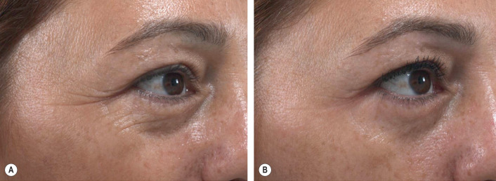
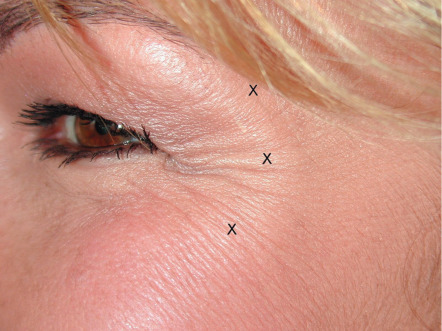
RT001 (Revance Therapeutics, Inc.) is the first topical formulation of BoNT-A and it combines purified 150 kDa BoNT-A protein with a novel peptide, allowing for transdermal penetration. RT001 was evaluated in five dose-escalation studies as part of a phase II clinical program , along with initial reports by Brandt and Glogau showing excellent efficacy for the treatment of lateral canthal rhytides ( Fig. 159.2 ). These investigations observed mild and transient side effects that did not appear to be dose-dependent and there was no evidence of diffusion to adjacent muscles, systemic spread, or safety issues. Topical BoNT-A may offer opportunities to treat anatomical areas that are difficult to manage or particularly painful to inject, such as palmar hyperhidrosis. Although phase III clinical trials of RT001 did not meet clinical endpoints, it is still under investigation. Another topical BoNT-A preparation, ANT-1207 gel, has successfully completed phase II clinical trials .
Initial studies of RT002, an injectable form of RT001, demonstrated great promise, with a long-lasting duration of effect and significantly less diffusion than onaA . Early results from a phase I/II trial of 48 patients with moderate-to-severe glabellar rhytides demonstrated a median duration of effect of 7.3 months and minimal side effects . Pivotal phase II clinical trials have been completed , and phase III trials of daxibotulinumtoxinA (RT002) are currently underway.
A note about dosing
Most of the published information regarding the cosmetic uses of BoNT pertains to the use of onaA, followed in recent years by aboA and incoA. Data are scarce regarding other formulations of BoNT-A. Since most of our experience pertains to the use of onaA, the following discussion (including units and description of techniques) focuses on this formulation specifically. However, a conversion rate of 1 : 1 U may be considered for all 100-U product vials (incoA, Prosigne ® , and Neuronox ® ) and 1 : 2.5 U for aboA.
Cosmetic Use of Botulinum Toxin Type A
Hyperkinetic lines result from the repeated contraction of muscles perpendicular to the rhytides. Weakening or relaxing these muscles with BoNT-A can smooth horizontal lines on the forehead (from frontalis contraction), vertical lines in the glabellar region between the eyebrows (from corrugator contraction), horizontal creases across the bridge of the nose (from procerus contraction), “crow’s feet” and lateral lines along the lower eyelid (from lateral orbicularis oculi contraction), and perioral lines (from orbicularis oris contraction). Deep grooves or folds elsewhere that are exacerbated by muscle activity are also amenable to treatment. Patients 30–50 years of age may be most responsive to BoNT-A, because their rhytides are more likely to be caused by muscle activity rather than by the loss of skin elasticity that occurs with aging.
Injections for aesthetic indications may be given intramuscularly, subcutaneously or intradermally. We use intradermal injections, especially in the crow’s feet area, to minimize bruising. All of the usual precautions prior to any injection should be followed. The area may be chilled with ice before the injections to minimize any discomfort in sensitive individuals. Alternatively, a topical anesthetic can be applied 15 to 30 minutes prior to injection to minimize discomfort.
The effects of BoNT-A injections are usually apparent within a day or two and are obvious for 3 or 4 months, although they may last for 6 months or longer. There is a tendency for repeated injections to provide aesthetic improvement that lasts longer. Current research indicates no significant difference between BoNT-A formulations in terms of duration of effect ( ).
Glabellar Frown Lines
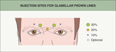
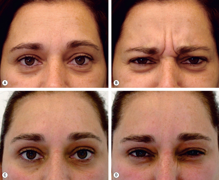
Frown lines in the glabellar region are caused by contraction of the corrugator supercilii and orbicularis oculi muscles, which move the brow medially, and the procerus and depressor supercilii muscles, which pull the brow inferiorly. Because the corrugator and procerus are used only to control facial expression, the goal of treatment should be to produce a significant weakening of these muscles. The treatment sites and doses should be individualized because the location, size and use of the frown muscles vary greatly between individuals. We currently use five injection sites when treating glabellar frown lines and vary the dosage depending on the individual brow ( Fig. 159.3 ; Table 159.2 ). The procedure effectively smooths the glabellar lines at rest and prevents their appearance when the patient attempts to frown, for an average of 3 to 4 months ( Fig. 159.4 ) ( ).
| USE OF onaA IN THE UPPER FACE: DOSING AND TECHNIQUES | |
|---|---|
| Treatment area | Injection sites and tips |
| Glabellar rhytides |
|
| Lateral canthal rhytides (crow’s feet) |
|
| Horizontal forehead rhytides |
|
| Hypertrophic orbicularis oculi muscle |
|
* Injection site most likely to cause ptosis.
† More than 12 cm between the temporal fusion lines (the slight prominence at the point where the temporalis fascia joins the skull) at the midforehead level.
Crow’s Feet
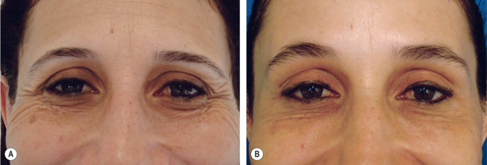
Contraction of the lateral fibers of the orbicularis oculi muscle produces “crow’s feet”, lines that radiate from the lateral canthus. Since forceful closure of the eyelids requires orbicularis contraction, the goal of treatment is to produce weakening just in the lateral orbital area, rather than a complete paralysis of the muscle. Because the orbicularis oculi is diffusely innervated, multiple injections are required to weaken broad areas of the muscle. While doses and sites vary among clinicians, we evenly distribute the BoNT-A into two to four separate injection sites ( Fig. 159.5 ; see Table 159.2 ). Results last approximately 3 months ( Fig. 159.6 ) ( ).
Horizontal Forehead Lines
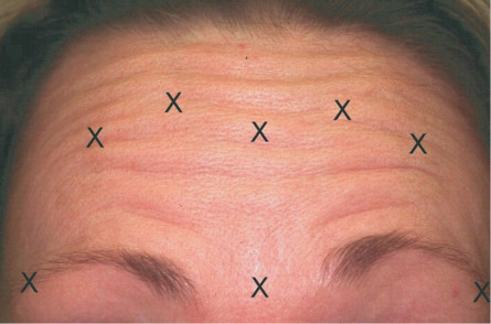
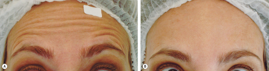
Deep horizontal creases in the forehead are produced by the repeated contraction of the frontalis muscle. This large, vertically oriented muscle is the brow elevator; it inserts superiorly into the galea aponeurotica and inferiorly into the skin of the brow. Unfortunately, weakening the frontalis muscle sufficiently to eliminate hyperkinetic forehead lines can result in undesired brow ptosis or a complete lack of expressiveness. Therefore, it is our practice to divide the BoNT-A amongst multiple sites distributed horizontally across the midforehead ( Fig. 159.7 ; see Table 159.2 ). The goal of treatment is to only soften, rather than completely eliminate, forehead lines. Ideally, the individual will still be able to elevate the eyebrows, albeit to a lesser extent, after treatment ( Fig. 159.8 ). We believe that the brow depressors should always be treated at the same time as the frontalis, and so we employ the technique described in the next section. Effects typically last from 3 to 6 months ( ).
Brow Lift and Shaping
Brow ptosis often occurs during aging, resulting in an angry, scowling expression. Notably, the shape and height of the eyebrows are determined by the opposing activity of the frontalis muscle, which elevates the brow, and the brow depressors. The medial brow depressors are the corrugator supercilii, the procerus, and the medial portion of the orbicularis oculi; the lateral depressor is the lateral portion of the orbicularis oculi (that is lateral to the temporal fusion line). Although we initially believed that brow lifts were a result of the inactivation of the brow depressors, subsequent research showed that central injections of 20–40 U of onaA into the glabella alone (with the most lateral injection at the midpupillary line) led to a dramatic lateral eyebrow elevation, followed by an entire brow lift that peaked 12 weeks after treatment, while too little (10 U) led to a brief fall in eyebrow position . We now believe that the change in eyebrow position after glabellar injection in women is due to diffusion of the toxin into the lower frontalis muscle, which causes an improved resting tone in the remainder of the muscle and in eyebrow position.
Hypertrophic Orbicularis Oculi
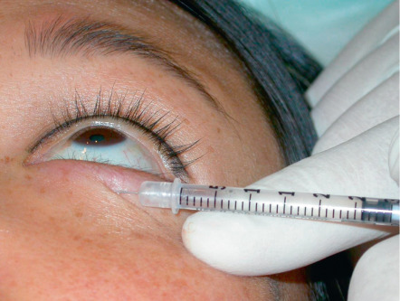
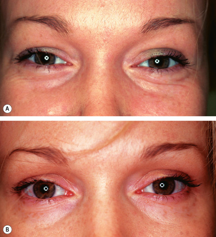
The pretarsal portion of the orbicularis oculi muscle is involved in the blink reflex. Contraction of the pretarsal orbicularis during smiling tends to decrease the size of the palpebral aperture. Hypertrophy of this muscle can give a “jelly roll” appearance to the lower eyelid, enough that some individuals may complain that they look overweight. We have found that injection of 2 U (or occasionally 4 U) of onaA into the lower pretarsal orbicularis ( Fig. 159.9 ) opens the palpebral aperture both at rest and during smiling ( Fig. 159.10 ; see Table 159.2 ) .
Midface Injections
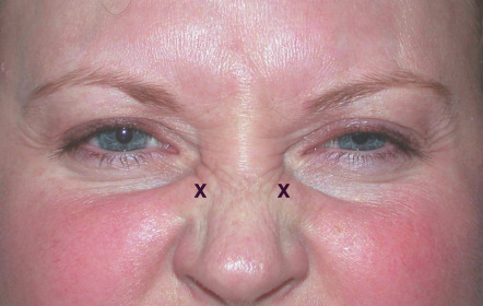
Contraction of the upper nasalis muscle causes fanning rhytides (“bunny lines”) at the nasal root ( Fig. 159.11 ). BoNT-A treatment weakens the upper nasalis and dramatically softens these lines. Bunny lines are typically treated in conjunction with the glabellar complex ( Table 159.3 ).
| USE OF onaA IN THE MIDFACE: DOSING AND TECHNIQUES | |
|---|---|
| Treatment area | Injection sites and tips |
| Bunny lines |
|
| Repeated nasal flare |
|
| Nasolabial folds |
|
| Nasal tip droop |
|
Related posts:


Stay updated, free articles. Join our Telegram channel

Full access? Get Clinical Tree


