Abstract
It is important for dermatologists to appreciate the human preoccupation with external beauty and aging. Having a better understanding of what constitutes beauty and how to evaluate the aging face helps us to better serve our patients as positive self-perception of physical appearance improves an individual’s quality of life. In this chapter, the physical characteristics that are viewed as signs of beauty will be discussed in addition to the concept of discrete facial fat compartments. Signs of chronologic aging and photoaging will be reviewed as well as ethnic and gender differences. A therapeutic approach to the aging face will be provided.
Keywords
beauty, aging, evaluation of beauty, facial fat compartments, photoaging, botulinum toxin type A (BoNT-A), soft tissue fillers, aging face
Introduction
“It is amazing how complete is the delusion that beauty is goodness.” ~Leo Tolstoy
Nancy Etcoff, in Survival of the Prettiest: the Science of Beauty , explains that beauty is a universal component of the human experience which promotes pleasure, rivets attention, and impels action that helps ensure survival of our genes .
Striving to be more beautiful is not a new phenomenon. Human preoccupation with beauty and a youthful appearance dates back as far as recorded history. Anthropologists have determined that over 40 000 years ago, red ochre paints were used in southern Africa for facial and body ornamentation . In 5 BC, the Roman author Ovid wrote The Art of Beauty, the first known book on beauty and cosmetics, detailing the use of black eye shadow made from wood ash and golden eye shadow made from saffron.
Few human traits are as ingrained as beauty. It is thought to be hardwired into our brains and can even be detected in infants. Studies have shown that by the age of 3 months, babies stare longer at attractive faces than those deemed to be less attractive. Also, 1-year-old toddlers will play longer with attractive dolls than with plain dolls . In functional magnetic resonance imaging studies where subjects were shown photographs of attractive men and women, viewing beautiful faces activated reward circuitry within the brain and dopamine pathways .
If examined from an evolutionary standpoint, this innate attraction to beauty certainly makes sense. Beauty has been interpreted in terms of reproductive potential as it provides an advantage in selecting or being selected as a mate for sexual reproduction. Attraction to beauty also makes sense from a disease perspective. It is said that “beauty is a burden that only the healthy can carry”. In other words, the sickly are by nature less attractive. Those with an ideal physique and youthful proportions are much more likely to be healthy and be able to reproduce. Again, by choosing an attractive healthy mate, one is increasing the odds that his or her genes will be passed on.
In one study, 20 subjects were asked to grade graduates from a 1920s high school yearbook on their attractiveness. When the actual lifespans of these graduates were determined, facial attractiveness correlated with increased lifespan. Correlations between beauty and health help to explain why, from an evolutionary standpoint, the notion of beauty may become an innate concept within a species .
Besides being considered a more ideal mate, attractiveness may also have a number of other benefits. Nancy Etcoff has stated that people tend to respond positively to good-looking people without expectation of reward, and, further, that good-looking people tend to be less penalized for everything from shoplifting to cheating at exams . In the same workplace, attractive people were found to earn up to 12% higher wages as compared to co-workers who were considered average-looking . Also, pharmaceutical representatives have been shown to have a 1.9% increase in sales for every unit increase in the Likert Scale for attractiveness. While the average height of a man in the US is 5′ 7″ (170.2 cm), the average height of Fortune 500 CEOs is 6′ 1″ (185.4 cm). Statistically, US Presidents have also been found to be significantly taller than their constituents. Lastly, even West Point graduates who had higher scores on an attractiveness scale rose to higher military ranks.
Evaluation of Beauty
Recognizing beauty and understanding its significance may be far easier than actually defining it . Anthropometry represents the quantitative measurement of facial features and an assessment of resultant ratios. It is based on proportional relationships of the face that are known as the neoclassical canons, which have been studied and revised for centuries. As originally proposed by Leonardo da Vinci, the ideal face can be divided horizontally into three equal parts; these parts are composed of the distances between: (1) the frontal hairline and the inferior edge of the frontal bone; (2) the supraorbital margin and the base of the nose; and (3) the base of the nose and the inferior aspect of the chin ( Fig. 152.1A ) . In general, mathematically idealized facial proportions translate into a symmetric, oval- or heart-shaped face, with prominent cheekbones, tapered jaw line, narrow nasal base and thin lips. These proportions, however, are largely based on facial features in Caucasian women and ethnic variations of these “ideal” proportions do exist.
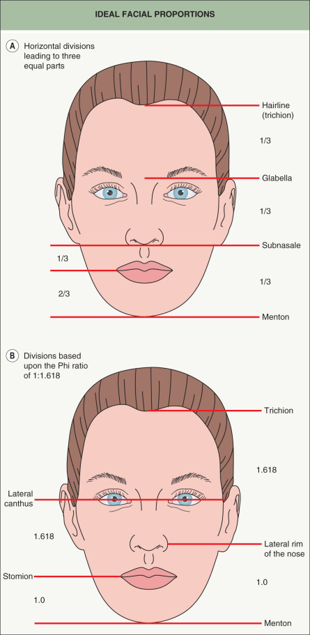
Another method for calculating beauty via mathematical proportions is the concept of “Phi”, or the golden ratio. A ratio of 1 : 1.618 was described by ancient Greeks as a mathematical method for calculation of the optimal proportions for all structures in nature ( Fig. 152.1B ). In fact, some authorities believe that the neoclassical canons (see above) are modifications of Phi. Phi represents the unique point on a line that divides the line into two lines in such a manner that the ratio of the smaller portion to the larger portion is the same as the ratio of the larger portion to the whole line ( Fig. 152.2 ). This ratio is a repeating number that is rounded off to 1 : 1.618. Of note, there are innumerable examples of the use of the Phi ratio in nature and art, including the Parthenon, the sculpture Venus de Milo, the shell of a nautilus, and the petals of a sunflower.

A triangle composed of lines with lengths in the Phi ratio is referred to as a golden triangle. When expanded, one can even create golden pentagons and golden decagons. Stephen Marquardt trademarked the “Phi mask”, a facial mask composed of proportions that incorporate the 1 : 1.618 ratio, to describe the mathematically ideal attractive face. Although the Phi mask has been applied to persons of all races and ethnicities, Marquardt recognized that ethnic variations may exist and has incorporated modifications for three different ethnic groups – Caucasian, Asian, and African.
Anatomic Basis for the Aging Appearance
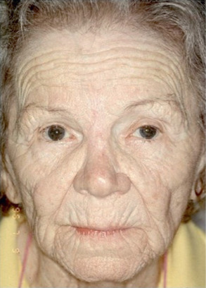
Knowledge of facial anatomy and anatomic changes that occur with aging is essential for properly evaluating and treating the aesthetic patient. With aging, both the golden ratio (Phi) and the neoclassical canons start to shift. The upper one-third of the face elongates due to recession of the frontal hairline and brow ptosis, while proportionately the middle third remains relatively stable (but may elongate with nasal tip ptosis). However, midface volume loss can occur as a result of fat redistribution and skeletal changes. The lower third of the face becomes shortened with perioral fat redistribution and significant resorption of the bony mandible.
The appearance of the aging face is multifactorial and does not appear to simply be a matter of sagging from the effect of years of gravitational forces pulling the face downwards. Rather, a complex ongoing alteration in proportions occurs as one ages. Notably, studies by Rohrich and Pessa have shown that there is not one large confluent layer of subcutaneous fat in the face but rather multiple distinct compartments that act independently from one another (see below, Volume changes ). For example, the malar fat pad in the mid cheek is composed of three separate compartments; as these compartments change in size, the ligaments separating the compartments become more evident and a smooth, rounded, youthful appearance transforms into the multi-contoured aged face.
After examining numerous photographs of patients as they aged, Lambros concluded that there is actually very little positional change in the midface region. Despite the common perception that the face drops inferiorly with age, he noted that the eyelid–cheek margin did not change with advancing years, nor did facial nevi of the midface descend with age. Just as individual water molecules simply move up and down rather than actually traveling with waves at sea, the wave of downward motion “seen” in an aging face may simply represent shifts in subcutaneous tissue.
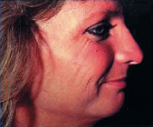
Rhytides also play a key role in producing an aged appearance. The underlying causes of rhytides are multiple and include ultraviolet radiation-induced damage, loss of skin elasticity, repetitive movement of underlying facial musculature, changes in volume from fat redistribution, and bone as well as cartilage resorption ( Fig. 152.3 ).
Photoaging
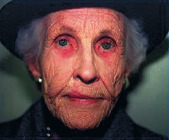
Cumulative exposure to sunlight is a major factor in aging skin. Clinical signs of cutaneous photoaging include: rhytides, lentigines, keratoses, telangiectasias, loss of translucency, loss of elasticity, and a sallow color . The Fitzpatrick Scale of Skin Phototypes (see Table 0.6 ) serves as a good indicator of the potential for developing signs of photoaging.
A simple systematic classification of patient photoaging types (types I through IV) was developed by Dr Richard Glogau ( Table 152.1 ) . However, the generalizations outlined in this table are influenced by the amount of cumulative sun exposure and usually apply at a later age and to a lesser degree in those with more darkly pigmented skin. Type I refers to younger patients with no rhytides, even when the face is animated by talking or expression. Type II refers to patients with “wrinkles in motion”, who demonstrate the influence the underlying musculature has on the skin.
Related posts:


Stay updated, free articles. Join our Telegram channel

Full access? Get Clinical Tree


