Axillary and Inguinal Creases
OVERVIEW
The word “intertrigo” comes from the Latin, terere, to rub, a catch-all word for the more descriptive term “intertriginous dermatitis.” Thus, when used in its more inclusive sense, intertrigo refers to an inflammatory condition of skinfolds. Intertriginous areas are places where skin is subject to the friction of repeated movement, chafing, obesity, increased local heat, and maceration from retained moisture and from topical applications, all of which serve to provide an excellent environment for potential irritant, fungal, as well as bacterial complications. Both the axillae and inguinal creases are “hot spots” for intertrigo.
Besides the inguinal creases and axilla, the body has other intertriginous appositional places such as the inframammary areas (see Fig. 11-53), finger webs (see Fig. 14-28), toe webs, perineum, and abdominal creases, all represent two opposing surfaces that are incessantly in intimate contact.
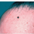
The potassium hydroxide (KOH) test, Gram stain, and bacterial culture are useful to exclude primary or secondary infection and to guide therapy. The sun-shaded axillae and inguinal creases are unlikely to absorb much in the way of ultraviolet exposure and are generally spared many skin cancers, although various benign skin growths such as skin tags and melanocytic nevi are commonly found there.
Since both the inguinal and axillary creases share many of the same cutaneous disorders, they will be discussed together in this chapter.
Irritant Intertrigo
Irritant intertrigo of the axillary vaults and inguinal creases is a very common problem. Both areas are subject to a host of irritants such as soap, shaving, deodorants, etc. Intertrigo is particularly difficult for patients with atopic dermatitis who already have an inherent skin sensitivity and defective skin barrier function.
Distinguishing Features
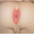
Begins as a mild erythema followed by erythematous well-demarcated patches or plaques that oppose each other on either side of the skinfolds like a mirror image (Fig. 12-1)
An atopic history is frequently elicited
Often pruritic; may progress to erosions, oozing, exudation, and painful fissures within the plaques or develop a secondary infection with coagulase-positive Staphylococcus aureus or Candida species (see below)
Diagnosis
Clinical
Exclusion of other diagnoses e.g., inverse psoriasis, seborrheic dermatitis, as well as bacterial or fungal infection
Negative potassium hydroxide (KOH) and/or fungal culture, although such infections may be secondary phenomena
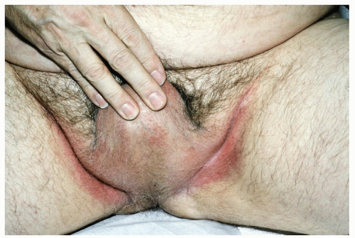 Figure 12-1 Irritant intertrigo. A moist, red, often macerated rash of the groin in an overweight person is characteristic. This condition is frequently confused with tinea cruris. |
Management
Prevention
Promote drying, aeration, and skin-to-skin friction (e.g., air conditioning, fans, hair dryer set on “cool”)
Zeasorb powder
Nonrestrictive clothing
Weight loss, if indicated
Gentle underarm shaving techniques
Avoidance of oily or irritant ointments or cosmetics
Treatment
Burow solution compresses to exudative, oozing areas
The lowest potency nonfluorinated topical steroids are used to avoid atrophy and striae. To achieve rapid improvement, treatment may be initiated with a higher-potency (class 5) steroid that is used for several days before it is changed to a lower-potency (class 6 or 7) agent.
It should be noted that overuse and/or continuous application of topical steroids in the intertriginous axillary and inguinal creases may cause stretch marks (striae) and marked thinning of the skin. For longer-term use and maintenance, the following agents can be applied without concern regarding steroid atrophy in intertriginous locations:
Tacrolimus 0.1% ointment (Protopic) once or twice daily
Pimecrolimus 1% cream (Elidel) once or twice daily
Crisaborole 2% ointment (Eucrisa) can be applied without concern regarding steroid atrophy in intertriginous locations. Approved primarily for the treatment of eczema in children >3 months of age, it can also be an effective tool for treating intertrigo and inverse psoriasis (see below)
Allergic Contact Dermatitis
Allergic contact dermatitis (ACD) may result from reaction to agents such as a dry-cleaning solvent (e.g., formaldehyde), topical medications, as well as ingredients in deodorants and antiperspirants.
Distinguishing Features
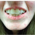
Mild erythema initially, followed by erythematous eczematous plaques and/or vesico-bullae (Fig. 12-2)
Pruritus may be severe
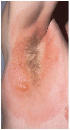 Figure 12-2 Allergic contact dermatitis. Acute blistering contact dermatitis determined to be secondary to formaldehyde in dry cleaning fluid (note sparing of the axillary vault due to less contact from his garment in this location). |
Diagnosis
History of contactant
Patch testing, if necessary, is used to confirm and identify a suspected allergen (see Appendix: Diagnostic and Therapeutic Techniques; Fig. A-12, A and B)
Management
Patients should be advised to avoid the offending agent or to minimize contact with it
Low-potency topical steroids
Inverse Psoriasis
When psoriatic lesions occur primarily in the intertriginous areas such as the axillae, inframammary, perineal, and inguinal creases, it is referred to as inverse psoriasis. Inverse psoriasis may also involve the penis and gluteal cleft (see Figs. 16-18 and 17-14).
Because of the moist nature of the skinfolds, the appearance of the psoriatic plaques is somewhat different than that seen on the elbows and knees—they generally do not have the typical silvery scale in areas of skin to skin contact where two opposing surfaces do not allow scale to build up.
There also may be fissuring in the depth of the skin creases. Psoriasis in this location is often difficult to distinguish from irritant intertrigo, atopic dermatitis, and candidiasis.
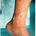
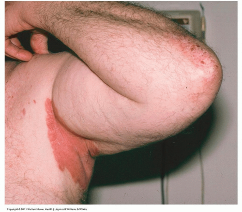 Figure 12-3 Inverse psoriasis. This is a well-defined pink plaque. Due to occlusion, there is less scale that is typically seen in psoriasis elsewhere such as is present on this man’s elbow. |
Distinguishing Features
The deep pink to red color and well-defined borders characteristic of psoriasis may be obvious; however, they generally lack scale (Figs. 12-3 and 12-4)
May be pruritic
Fissures may occur
Diagnosis
Clinical
Psoriasis may be present elsewhere on the body
Negative KOH and/or fungal culture
Management
Low-potency, nonfluorinated topical steroids (class 6 or 7) or, if necessary, a higher-potency (class 5) steroid that is used for several days before changing to a lower-potency agent
Stronger topical steroids need to be used with care and only for a few days at a time
Systemic agents are rarely required for limited flexural psoriasis, and phototherapy is ineffective because the skinfolds are hidden from ultraviolet light exposure
Vitamin D-like compounds such as calcipotriol Dovonex and Vectical cream or ointment may be used primarily or in rotation with a mild topical steroid
Calcineurin inhibitors, tacrolimus 0.1% ointment (Protopic), or pimecrolimus 1% cream (Elidel) is applied once or twice daily for longer-term use
Different topical medications are used together or in rotation for best effect or to minimize side effects
Crisaborole 2% ointment (Eucrisa) can be applied without concern regarding steroid atrophy in intertriginous locations
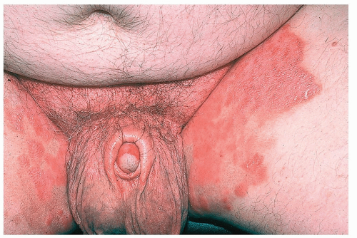 Figure 12-4 Inverse psoriasis. Note involvement of the scrotum, foreskin, and the glans. This eruption can be easily confused with tinea cruris. |
Cutaneous Candidiasis
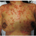
Cutaneous candidiasis is characterized by an infection with Candida species. Candida is a common secondary and sometimes primary cause of intertrigo in elderly, diabetic, or immunocompromised patients. Most cases occur in skinfolds, where occlusion by clothing produces warm, moist conditions. Candidal infection of the skin under the breasts occurs when such areas become macerated under pendulous breasts (see also Fig. 11-55).
Distinguishing Features
“Beefy red” lesions
Satellite pustules may be seen beyond the border of the plaques (Figs. 12-5 and 12-6)
Maceration and fissures may be present
Soreness and/or pruritus
Diagnosis
Positive KOH examination for budding yeast or positive culture for Candida species
Management
For acute candidal intertrigo, Burow solution compresses may be applied to exudative, oozing areas
The skin should be patted dry or dried with a hair dryer set on “cool”
Topical ketoconazole, clotrimazole, or miconazole twice daily
Patients with extensive infection may require the addition of oral fluconazole (Diflucan), itraconazole (Sporanox), or ketoconazole
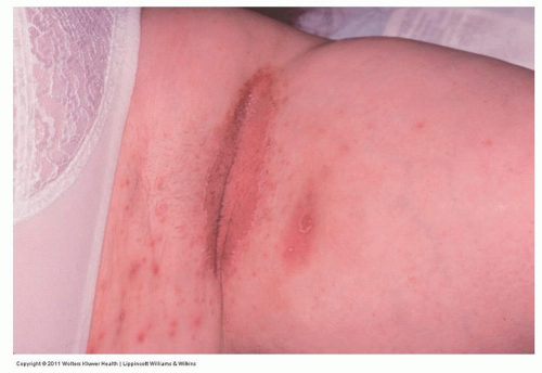 Figure 12-5 Candidiasis, axillary. Erythema of the axillary crease and “satellite” pustules. |
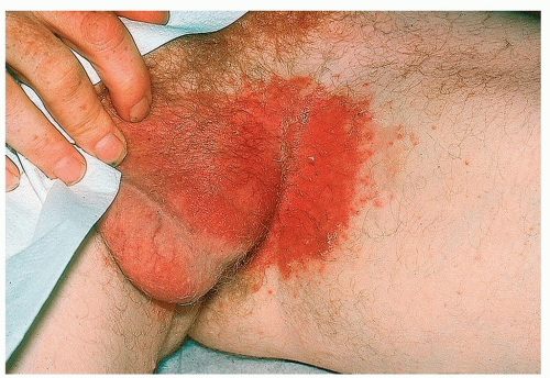 Figure 12-6 Candidiasis, inguinal. Note beefy red color and a few “satellite” pustules. |
Tinea Cruris
Tinea cruris (TC), also known as “jock itch,” is a common infection of the upper inner thighs and crural area, most often occurring in postpubertal male patients. It is generally caused by the dermatophyte Trichophyton rubrum. In contrast to candidiasis and lichen simplex chronicus, it generally spares the scrotum.
Related posts:
Stay updated, free articles. Join our Telegram channel

Full access? Get Clinical Tree


