Abstract
Autoantibody testing is integral to the clinical evaluation of suspected autoimmune connective tissue disease as it aids in disease classification and can sometimes be used to follow disease activity. In addition, autoantibodies can be used in the evaluation of systemic vasculitides. Certain autoantibodies have significant disease specificity and thus can be of great diagnostic value: anti-dsDNA and anti-Smith (Sm) for systemic lupus erythematosus (SLE); anti-Mi-2 for classic dermatomyositis; anti-Jo-1 for antisynthetase syndrome; anti-topoisomerase-1 (Scl-70), anti-RNA polymerase III, and anti-centromere for different clinical forms of systemic sclerosis; and cytoplasmic antineutrophil cytoplasmic antibodies (c-ANCA) for granulomatosis with polyangiitis. However, many autoantibodies fall into the disease-nonspecific category. The principles, techniques, and clinical uses of autoantibody testing are reviewed.
Keywords
autoantibodies, antinuclear antibody, ANA, autoimmune connective tissue disease, systemic lupus erythematosus, ANCA, c-ANCA, p-ANCA, Scl-70, anti-topoisomerase-1 antibody, anti-centromere antibody, antisynthetase syndrome
- ▪
Autoantibodies can be of significant value in the diagnosis, management, and prognosis of autoimmune connective tissue diseases (AI-CTDs), but their interpretation depends on the type of autoantibody and the specific AI-CTD
- ▪
The classic ANA assay remains the entrée into the world of AI-CTD serology, and understanding its limitations is critical to clinical decision making
- ▪
The evolution of clinical immunology laboratory technology has altered the significance of some of the original clinical–serologic correlations reported at the time of discovery of the autoantibodies. Basic understanding of key technical issues related to current laboratory methodologies is therefore important
- ▪
There has been a trend toward adopting solid-phase immunoassay techniques (e.g. ELISA) to detect many autoantibodies. For several autoantibodies, including anti-SSA/Ro and anti-double-stranded (ds) DNA antibodies, this has resulted in some decrease in disease specificity
- ▪
Certain autoantibodies have significant disease specificity and thus can be of great diagnostic value: anti-dsDNA and anti-Smith (Sm) for systemic lupus erythematosus (SLE); anti-Mi-2 for classic dermatomyositis; anti-Jo-1 for antisynthetase syndrome; anti-topoisomerase-1 (Scl-70), anti-RNA polymerase III, and anti-centromere for different clinical forms of systemic sclerosis; and cytoplasmic antineutrophil cytoplasmic antibodies (c-ANCA) for granulomatosis with polyangiitis. However, most autoantibodies fall into the disease-nonspecific category
- ▪
The absolute blood levels of a few autoantibodies can correlate positively with underlying autoimmune disease activity (e.g. anti-dsDNA in SLE, c-ANCA in granulomatosis with polyangiitis), but in most cases the titer does not correlate with disease activity
Introduction
Autoimmune connective tissue diseases (AI-CTDs) represent polygenic clinical disorders that have heterogeneous and overlapping clinical features. Sometimes they are referred to as “autoimmune rheumatic diseases”. These disorders are characterized by an immune dysregulation that includes the production of antibodies to self-antigens, termed autoantibodies (aAb). The aAb in patients with AI-CTD often target structures vital to cellular metabolism and division.
When the diagnosis of an AI-CTD is being considered, the most commonly requested laboratory test is measurement of antinuclear antibodies (ANA). ANA are commonly defined as aAb that target primarily nuclear components, including DNA or small nuclear ribonucleoproteins (snRNP), and are detected by the fluorescent antinuclear antibody (FANA) test. Elevated titers of ANA are seen in multiple AI-CTD, from systemic lupus erythematosus (SLE) to systemic sclerosis (SSc) and dermatomyositis (DM). These aAb reflect basic inflammatory events in tissue, but rarely carry pathogenic potential (the exception being ANCA). ANCA target cytoplasmic structures and assist in the diagnosis of systemic vasculitides (see Ch. 24 ).
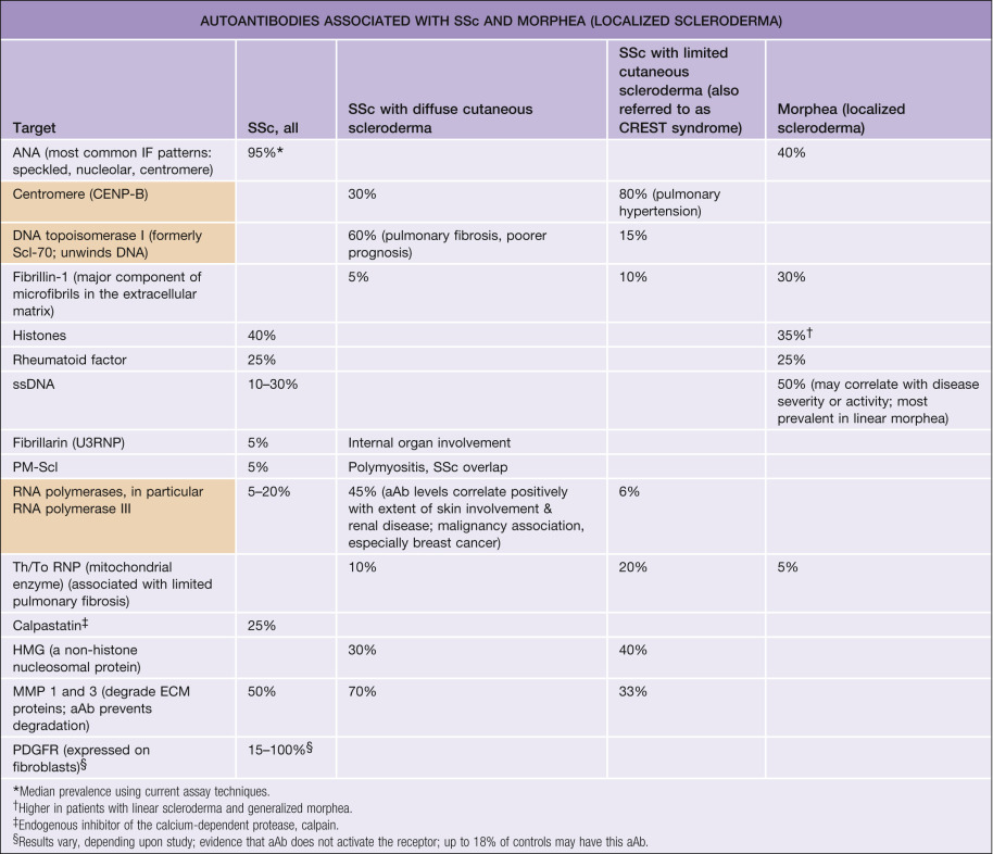
If used appropriately, clinical laboratory testing for ANA can be useful in the diagnosis and management of AI-CTD patients . In order to maximize the clinical utility of aAb testing in this setting, familiarity with the serologic tests currently used to identify these aAb, as well as the disease associations of the various aAb, is helpful. This chapter focuses on these latter two aspects, while Chapter 41 , Chapter 42 , Chapter 43 , Chapter 44 , Chapter 45 contain an in-depth discussion of the various AI-CTDs. Therefore, the molecular identity of the various autoantigens and their clinical associations are summarized in table form (see Tables 40.2–40.4 ). An effort will also be made to illustrate the often complex relationships that can exist between the presence and relative amounts of these aAb, disease diagnosis, and prognosis. In addition, the basic principles of clinical utility as related to the various aAb laboratory tests (e.g. sensitivity, specificity) will be briefly addressed. The related subjects of circulating immune complexes and cryoglobulins are discussed in Chapter 4 , Chapter 23 , Chapter 24 .
| AUTOANTIBODIES ASSOCIATED WITH LUPUS ERYTHEMATOSUS | |||
|---|---|---|---|
| Target † | Median prevalence * | Molecular specificity | Clinical associations |
| High specificity for SLE | |||
| dsDNA | 60% | Double-stranded (native) DNA | LE nephritis and monitoring activity of nephritis |
| Sm | 10–30% of Caucasians; 30–40% of Asians and African-Americans | Splicesome RNP (ribonucleoprotein particles involved in splicing pre-mRNA) | |
| rRNP | 7–15%; 40% of Asians | Ribosomal P proteins (proteins involved in ribosome function) | Neuropsychiatric LE |
| Low specificity for SLE | |||
| ANA (most common IF patterns: homogeneous, peripheral) | 99% | ||
| ssDNA | 70% | Denatured DNA | Possible risk for SLE in DLE patients; also seen in RA, DM/PM, MCTD, SSc, SjS, morphea |
| C1q | 60% | C1q component of complement | Severe SLE, hypocomplementemic urticarial vasculitis syndrome |
| PCNA | 50% | A component of multiprotein complexes involved in cellular proliferation | – |
| U1RNP | 50% | Splicesome RNP | Overlapping features with other AI-CTDs; MCTD (100%) |
| SSA/Ro | 50% | hYRNP (quality control function for misfolded RNA molecules) | SCLE (75–90%), neonatal LE/congenital heart block (99%), SCLE–SjS overlap, primary SjS (70%); associated with vasculitis |
| SSB/La | 20% | hYRNP | SCLE (30–40%), SCLE–SjS overlap, primary SjS (40%); occurs in conjunction with SSA/Ro |
| Cardiolipin | 50% | Cardiolipin, a negatively charged phospholipid | Recurrent spontaneous abortions, thrombocytopenia, and hypercoagulable state in SLE (cutaneous manifestations include livedo reticularis, leg ulcers, acral infarction/ulceration, hemorrhagic cutaneous necrosis); similar associations in primary antiphospholipid antibody syndrome; clinical manifestations have strongest association with IgG class of anti-cardiolipin |
| β2 glycoprotein I | 25% | An important cofactor for cardiolipin in cardiolipin aAb assays | Relatively high risk of thrombosis in SLE and primary antiphospholipid antibody syndrome |
| Histones | 40% | Histones | Drug-induced SLE; also RA, SLE, and SSc with pulmonary fibrosis (in conjunction with other aAb) |
| Rheumatoid factor | 25% | Fc portion of IgG | Nonspecific |
| Ku | 10% | DNA end-binding repair protein complex | Overlap with other AI-CTDs such as DM/PM, SSc |
| Alpha-fodrin | 10% | An actin-binding protein found at the periphery of chromaffin cells that may be involved in secretion | SjS |
* Based on most common assay techniques currently employed in clinical immunology laboratories. Note that these figures represent the authors’ best estimates based on most recently published data.
† Listed in decreasing order of prevalence within categories.
| AUTOANTIBODIES ENCOUNTERED IN THE IDIOPATHIC INFLAMMATORY DERMATOMYOPATHIES | |||
|---|---|---|---|
| Target | Median prevalence * | Molecular specificity | Clinical association |
| High specificity for DM/PM | |||
| p155 | 80% (clinically amyopathic); 20–30% (classic) | Transcriptional intermediary factor 1 gamma (TIF1-γ) | Clinically amyopathic DM; in adult-onset classic DM, increased risk of malignancy; extensive cutaneous involvement; mucocutaneous findings include palatal erythema (“ovoid patches”), psoriasiform lesions, and hypopigmented and telangiectatic (“red on white”) patches |
| Mi-2 | 15% | Helicase nuclear proteins | Gottron papules/sign, shawl sign, cuticular telangiectasias, cuticular overgrowth/dystrophy |
| Jo-1 | 20% | Histidyl tRNA synthetase | Antisynthetase syndrome |
| PL-7 | 5% | Threonyl tRNA synthetase | Antisynthetase syndrome |
| PL-12 | 3% | Alanyl tRNA synthetase | Antisynthetase syndrome |
| OJ | Rare | Isoleucyl tRNA synthetase | Antisynthetase syndrome |
| EJ | Rare | Glycyl tRNA synthetase | Antisynthetase syndrome, possibly increased frequency of skin changes |
| SRP | 5% | Signal recognition particle (intracytoplasmic protein translocation) | Fulminant DM/PM, cardiac involvement |
| Fer | Rare | Elongation factor 1-α | − |
| Mas | Rare | Small RNA | − |
| MDA5/CADM-140 | 10–15% of Caucasians; 10–45% of adult Asians; 5 (in the UK) –35 (in Japan)% of juveniles | Melanoma differentiation-associated protein 5 (MDA5)/IFN induced with helicase C domain protein 1 (IFIH1) | Clinically amyopathic DM, rapidly progressive interstitial lung disease, cutaneous ulcerations with associated vasculopathy, tender palmar papules, oral pain and ulceration |
| NXP-2 | 1–15% of adults; 20–25% of juveniles | Nuclear matrix protein (MORC family CW-type zinc finger 3 [MORC3]) | In adults, associated with malignancy, subcutaneous edema, and calcinosis; in juveniles, associated with more severe muscle disease and calcinosis |
| Low specificity for DM/PM | |||
| ANA (most common IF patterns: speckled, nucleolar) | 40% | Clinically amyopathic DM (65%) | |
| ssDNA | 40% | Single-stranded DNA | SLE, SSc, morphea |
| PM-Scl (PM-1) | 10% | Ribosomal RNA processing enzyme | Overlap with SSc |
| SSA/Ro (especially 52 kDa Ro) | 15% | hYRNP | Overlap with SjS, SCLE, neonatal LE/CHB, SLE |
| U1RNP | 10% | Splicesome RNP | Overlap with other AI-CTDs |
| Ku | 3% | DNA end-binding repair protein complex | Overlap with SSc |
| U2RNP | 1% | Splicesome RNP | Overlap with SSc |
Chapter Organization
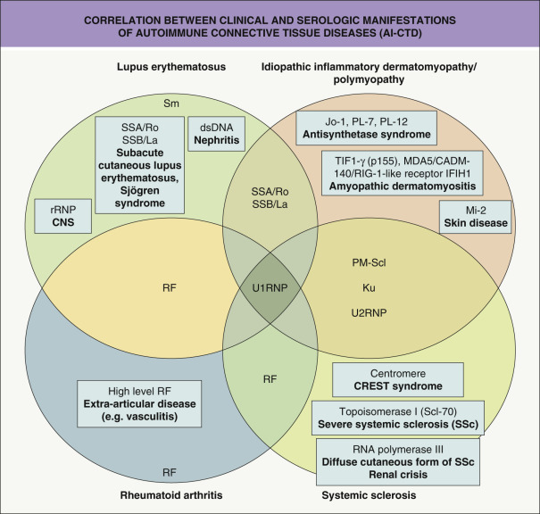
Although the various AI-CTDs are distinct clinical entities, they can have features in common. Consequently, each AI-CTD has aAb specific to that particular disorder as well as other aAb that are shared with the other diseases. In addition, some patients can display overlapping clinical features of several different AI-CTDs and, as a result, an overlap in the pattern of aAb production ( Fig. 40.1 ).
This chapter is organized based upon two major considerations in the use of aAb testing:
- •
historical and technical aspects of ANA assays
- •
aAb associated with AI-CTDs that have cutaneous manifestations.
Historical Perspective
One can better appreciate the evolution of thought that has occurred in this area by considering the change in methodology used to identify and measure these aAb over the past 60 years . In 1948, the bone marrow from a patient with SLE was noted to contain polymorphonuclear neutrophils that had phagocytosed nuclear material from degenerated cells. This became known as the LE cell phenomenon and occurred in the presence of antibody to DNA. The presence of these cells became an indicator of SLE. Approximately 10 years later, it was shown that indirect immunofluorescence could be used to detect ANA (i.e. the FANA test), providing a more sensitive assay for SLE.
Another decade later, Tan and co-workers employed the Ouchterlony double immunodiffusion technique to identify and define precipitating serum aAb that reacted with saline extractable nuclear antigens (ENA) such as nuclear ribonucleoprotein (nRNP), Sm, SSA/Ro, and SSB/La . Unfortunately, the Ouchterlony double immunodiffusion technique is time-consuming and expensive. However, a more efficient adaptation of the double immunodiffusion technique, counterimmunoelectrophoresis, can be used as an alternative to the less expensive solid-phase immunoassays (e.g. e nzyme- l inked i mmuno s orbent a ssay [ELISA]) that can be less specific (see below) .
In 1979, the molecularly and genetically defined ANA era began as the result of work by Lerner and Steitz, who employed immunodiffusion and Western blotting to define the molecular identities of the nRNP and Ro:La families of ribonucleoprotein autoantigens. These observations generated the notion that the various AI-CTD aAb might be more efficiently and cost-effectively assayed by using purified and/or recombinant forms of the various autoantigens in solid-phase immunoassays such as the ELISA. While less expensive and more convenient, there are a number of drawbacks to employing the ELISA and related immunoassay techniques to detect ANA such as anti-SSA/Ro and anti-dsDNA aAb, in particular increased sensitivity and decreased specificity. Newer techniques (e.g. solid-phase assays, proteome microarrays, Luminex xMAP ® technology) are now being used more frequently, allowing for the simultaneous profiling of all relevant aAb in a given patient. Understanding the actual immunochemical techniques used to detect and quantify the various types of ANA can be helpful to the clinician, but is beyond the scope of this chapter. The reader is referred to a review by Griesmacher and Peichl .
FANA: the Classic ANA Assay
Although there is debate about its role in the diagnosis and management of AI-CTD , the classic ANA indirect immunofluorescence assay (i.e. FANA) is still the most clinically efficient screening test for systemic autoimmune disorders such as SLE . A number of potential pitfalls must be considered when interpreting the results of the ANA assay ( Table 40.1 ). The following discussion will focus on the key aspects of interpreting ANA test results.
| MAJOR ISSUES RELATED TO THE INTERPRETATION OF RESULTS OF ANTINUCLEAR ANTIBODY (ANA) ASSAYS |
|
Importance of Technical Aspects of the ANA Assay
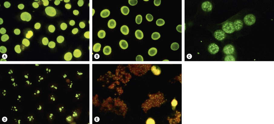
As the name implies, the ANA assay identifies antibodies present in serum that bind to autoantigens present in the nuclei (or cytoplasm) of mammalian cells. Both the pattern and the titer are reported , with the former consisting of morphological descriptors that reflect the localization of the autoantigen. The reported titer represents the last dilution at which the ANA pattern is detectable, and a titer of 1 : 40 or greater is considered “positive” (see below). The current version of the ANA assay used in almost all clinical laboratories employs a human tumor cell line such as Hep-2 for the nucleated cell substrate. aAb are detected with a fluorochrome-conjugated antiserum that is specific for human immunoglobulin (the aAb) that is bound to nuclei in the cell substrate ( Fig. 40.2 ). Earlier versions of the ANA assay utilized rodent cell lines and the latter lacked some of the autoantigens present in human cell nuclei (e.g. SSA/Ro). Thus, sera from some SLE patients could be negative when assayed on rodent cells, especially if the predominant aAb were anti-SSA/Ro (i.e. “ANA-negative SLE”). This occurred in up to 15% of certain SLE patient populations, particularly those enriched in anti-SSA/Ro aAb-associated disorders such as subacute cutaneous LE (SCLE) and Sjögren syndrome (SjS). Nowadays, because of the use of human Hep-2 cells, only ~1–2% of SLE patients are ANA-negative. Thus, “ANA-negative SLE” is predominantly an historical phenomenon.
An important caveat with regard to the FANA test is that determination of the titer (which reflects the serial serum dilutions necessary for fluorescence to disappear) depends upon subjective interpretation by a laboratory technician. Consequently, a single serum specimen can have an ANA titer result that varies within a two-tube dilution range. In other words, a serum sample reported as having an ANA titer of 1 : 320 might be read as 1 : 160 or 1 : 640 upon repeat testing in the same laboratory within the same timeframe. Although an attempt has been made by the World Health Organization to standardize reporting of ANA results with an international unit system (e.g. 1 IU rather than a titer of 1 : 160), many clinical laboratories in the US continue to report ANA results using a titer system. More recently, automated systems have been introduced in an attempt to decrease variability and increase cost-effectiveness; however, no pattern is reported and sometimes the result is simply positive or negative with no titer provided .
“Normal” Versus “Abnormal” ANA Values
The ANA titer that is considered to be abnormal can vary significantly, depending upon how the assay is performed and interpreted. The commercial ANA kits that are used most commonly in laboratories today usually indicate that an ANA titer of 1 : 40 is considered abnormal, presumably because accepting such relatively low levels of ANA as being abnormal retains a high degree of sensitivity of the ANA assay in detecting systemic AI-CTD. However, using such low ANA titer cut-offs creates a lot of positive, but clinically insignificant, ANA test results, i.e. it reduces specificity. A number of studies that have compared ANA results in SLE populations with those in normal control populations have indicated that a titer of <1 : 160, using a human tumor cell line substrate, has little clinical utility. For example, in one report based upon 15 international laboratories, the ANA positivity rate in a population of healthy individuals (ages 20–60 years) was 13.3% at 1 : 80, 5.0% at 1 : 160, and 3.3% at 1 : 320 . Of note, the elderly, relatives of those with SLE, and patients with other autoimmune disorders (e.g. autoimmune thyroid disease), as well as healthy individuals, may have abnormally elevated ANA titers.
Related posts:


Stay updated, free articles. Join our Telegram channel

Full access? Get Clinical Tree


