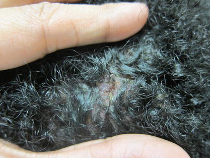
Figure 4.1
Acne keloidalis. Hyperpigmented follicular papules and pustules, along with hyperpigmented firm keloidal nodules, are present on the occipital scalp with associated areas of patchy alopecia

Figure 4.2
Acne keloidalis. Close up. Upon separate of the hair, erythematous follicular clustered papules with pustular drainage on the occipital scalp with patchy alopecia
Clinical Differential Diagnosis
This patient’s presentation was most consistent with acne keloidalis (AK). The differential diagnosis for AK includes acne vulgaris, folliculitis decalvans, dissecting cellulitis, and hidradenitis suppurativa. However the chronic and progressive nature of symptoms in an African American male, the exclusivity of his rash to the posterior neck, and the presence of keloidal plaques without evidence of comedones or signs of infection, made each of these diagnoses unlikely (Huggins 2014).
Histopathology
This disease entity is diagnosed clinically and biopsy is not necessary for pathologic confirmation. However if performed, histopathologic examination would reveal perifollicular inflammation with a neutrophilic and/or lymphocytic infiltrate surrounding the hair follicle in early disease. During later stages, fibroplasia, absence of sebaceous glands, and naked hair shafts in the dermis are seen with associated granulomatous inflammation (Herzberg et al. 1990).
Diagnosis
Acne keloidalis (AK)
Case Treatment
The patient was informed of the diagnosis and counseled on the inflammatory nature of the disease. He was advised to avoid mechanical irritation including scratching or wearing tight fitting clothing, high-collared shirts and hats, as well as avoidance of close hair clipping and/or shaving of the neck. His large, keloidal plaques were injected with 10 mg/mL triamcinolone acetonide both for symptomatic relief and to help flatten the lesions. For treatment of the remaining smaller lesions, he was prescribed topical clobetasol propionate 0.05 % foam, which he was instructed to use twice daily for a 2-week off-and-on regimen. In adjunct, he was prescribed topical tretinoin 0.025 % cream on an every other nightly regimen, and he was told to advance to once nightly as tolerated. The patient expressed significant concern about the cosmetic appearance of his keloidal plaques. After discussion, he agreed to return to clinic in 4–6 weeks to assess response to the intra-lesional and topical therapies and to consider the option of continued treatment with serial intralesional injections vs. surgical excision.
Discussion
Acne keloidalis (AK), also known as folliculitis keloidalis nuchae, is almost exclusively a disease of skin of color, as it predominantly affects post-pubertal men of African and Hispanic descent. Within this population, the prevalence has been estimated to range between 1.3 and 16.3 % (Huggins 2014). There are few case reports in the literature on affected black women or caucasian males.
A misnomer, acne keloidalis, is neither associated with acne vulgaris nor with true keloids. It is a progressive and chronic inflammatory folliculitis that presents with persistent pink, erythematous, or red-brown 2–4 mm papules on the occipital scalp and posterior neck that may progress overtime to form coalescing fibrotic “keloid-like” nodules. There is a risk of secondary infection, with development of pustules or purulent drainage. If infection is severe, draining sinuses and abscesses may result. Because destruction of the follicular unit is central to the pathophysiology of the disease, patients may also suffer from an associated scarring alopecia. They may also have “tufted” hairs in which multiple hair shafts emerge from one follicular orifice.
Histologic exam correlates with the evolving clinical features of the disease. During the initial inflammatory stage, there is perifollicular inflammation and subsequent weakening of the perifollicular wall. This follicular inflammation correlates with the appearance of papules on examination (Herzberg et al. 1990). In this early stage of disease, patients may be asymptomatic or may experience varying degrees of pruritus (Huggins 2014). Eventually, as the wall critically weakens, the naked hair shaft is released into the dermis. Overtime, this foreign body reaction induces surrounding granulomatous inflammation with extensive hypertrophic scarring, which manifests as fibrotic keloidal-appearing lesions (Herzberg et al. 1990). It is notable, that keloids are a distinct clinical entity, and by histology, they are characterized by an overabundant proliferation of thick collagen fibers. In later stages of AK, patients may have more severe pruritus and pain although some patients may have subclinical disease that presents only with hair loss (Huggins 2014).
Stay updated, free articles. Join our Telegram channel

Full access? Get Clinical Tree


