Introduction
Patients with a burn injury are at risk for a variety of surgical complications. These complications can affect multiple organ systems and occur either due to the burn injury itself or secondary to iatrogenic factors during the complex care of these patients. Burns induce both complex local and systemic inflammatory responses that can contribute to multisystem organ dysfunction. They can also occur in the setting of trauma, requiring thorough assessment and expeditious management as per Advanced Trauma Life Support (ATLS) guidelines.
Patients with large burns (>20% of the total body surface area [TBSA]) develop increased capillary permeability and intravascular volume depletion, typically requiring a prolonged hospitalization for fluid resuscitation with numerous debridement and skin grafting procedures. These patients are at increased risk for numerous short- and long-term surgical complications affecting multiple organ systems ( Fig. 31.1 ).
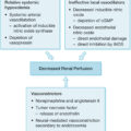
Schematic representation of the role of the gastrointestinal tract in multiorgan sepsis after cutaneous burn injury.
IL , Interleukin; TNF , tumor necrosis factor.
(From Gosain A, Gamelli RL. Role of the gastrointestinal tract in burn sepsis. J Burn Care Rehabil . 2005;26:85-91.)
In the acute resuscitation period, severe burn patients are at risk for extensive fluid shifts and labile hemodynamics. The burn injury contributes to capillary leak and intravascular volume depletion requiring large-volume fluid resuscitation. Therefore, patients typically require invasive lines, including central venous access and arterial lines, for resuscitation and hemodynamic monitoring. They may also require endotracheal intubation and an indwelling urinary catheter to monitor urine output as an assessment of resuscitation. Consequently, patients are at risk for catheter-related complications, such as catheter-associated infection and sepsis or distal limb ischemia, as well as ventilator-associated pneumonia. Underresuscitation can lead to decompensated burn shock and organ failure, whereas overresuscitation can induce pulmonary edema and abdominal compartment syndrome (ACS). The increased intraabdominal pressure (IAP) seen in ACS can necessitate decompressive laparotomy to prevent further end-organ damage.
Severe burn patients are also at increased risk for numerous other gastrointestinal (GI) complications in the acute and subacute periods. Occult systemic sepsis in burn patients is frequently secondary to a GI cause. Abdominal complications occur in approximately 1 of 20 burn patients with an associated mortality of 45%. The risk of these complications increases directly with burn size. Examples of these complications include stress gastritis and ulceration, acalculous cholecystitis, acute pancreatitis, superior mesenteric artery (SMA) syndrome, and ischemic enterocolitis, which can progress to bowel necrosis, perforation, and rapid clinical decline.
Management of burn patients requires a high index of clinical suspicion to mitigate delays in diagnosis and treatment of nonthermal complications. These include numerous surgical complications that can contribute to increased morbidity, prolonged hospitalization, sepsis, and even death. After the acute resuscitation period, septic shock is the predominant cause of death in burn patients. However, the early signs and symptoms of sepsis differ in burn patients and can be underrecognized given their hypermetabolic state, despite their increased risk due to their immunocompromised status. This chapter reviews the diagnosis and management of common surgical, nonthermal complications seen in burn patients.
Primary assessment
Prompt and appropriate initial management of burn injury is vital for optimizing patient outcomes. The primary survey of burn patients is identical to that of other traumas and should follow ATLS guidelines. The shocking appearance of burn injury may unduly shift attention away from other seemingly underwhelming injuries in patients with additional potentially life-threatening traumatic injuries, resulting in delays in diagnosis and worse outcomes. Except for respiratory compromise secondary to circumferential chest burns requiring escharotomies, the burn injury itself is typically not immediately life threatening. Primary assessment should therefore focus on airway, breathing, and circulation, followed by disability, and exposure/environmental exposure as per ATLS guidelines. The history of present illness can provide additional valuable information regarding the mechanism of injury, the likelihood of inhalation injury, and the probability of associated traumatic injuries.
Patients with burn injury warrant additional considerations when performing the primary survey. For instance, when assessing the airway, inhalation injury can induce burns above the vocal cords or edema that may worsen after fluid resuscitation. Inhalation injuries are common in burn patients, particularly in those with thermal injuries that occurred in enclosed areas and/or with prolonged extrication. Common signs of inhalation injury include erythema or edema of the oropharynx, carbonaceous sputum, cough, or stridor. Asphyxia can be seen with carbon monoxide poisoning, for which pulse oximetry is an unreliable source of oxygenation. Inhalation injury can progress rapidly over hours despite a normal initial chest radiograph or arterial blood gas. Smoke inhalation induces a multitude of physiologic changes, resulting in increased vascular permeability and pulmonary edema, infiltration by leukocytes, and bronchorrhea. The oropharynx should be carefully inspected and intubation or a surgical airway should be performed if there is concern regarding airway patency.
After the assessment of the airway, the primary survey should proceed with an evaluation of the patient’s breathing. Thoracic injuries, such as pneumothorax and hemothorax, should be managed with thoracostomy tubes, as they would be for any other blunt or penetrating traumatic injury. However, because burn wounds carry an increased risk of infection, thoracostomy tubes should be placed away from burned skin when possible to reduce the risk of infectious complications such as empyema. Circumferential full-thickness burns to the chest can impair respiratory mechanics, requiring escharotomy to release the constrictive eschar. Electrocautery can be used to incise the eschar to the level of the subcutaneous fat from the clavicle to the subcostal margin along the bilateral anterior axillary lines, followed by transverse incisions of the upper chest and/or upper abdomen to connect them. ,
Circulation should subsequently be assessed. Intravenous access should be established. Profound hypovolemia is uncommon immediately after a burn injury. Severe hypotension should raise suspicion for delayed presentation, cardiogenic dysfunction, or hemorrhage secondary to traumatic injury. Blunt cardiac trauma can result in pericardial tamponade, which should be assessed using focused assessment with sonography for trauma (FAST) examination and managed with pericardiocentesis or pericardial window. Myocardial dysfunction can also be seen secondary to electrical injuries, which should be evaluated with an electrocardiogram and managed accordingly. Circumferential full-thickness burns can function as a tourniquet, limiting perfusion particularly after resuscitation. In the event of decreased perfusion, the tissue should be released with escharotomies.
Disability should be assessed using the Glasgow Coma Scale. If a patient with a burn injury is not alert and oriented, suspicion should be raised for causes such as traumatic injury resulting in hypovolemia, carbon monoxide poisoning, hypoxia, or ingestion of substances.
Complete exposure of the patient should be obtained to allow for the estimation of TBSA and to assess for any concomitant injuries. Burn patients should then be promptly covered and warmed to prevent hypothermia. Fluid resuscitation and analgesia should be administered as indicated following the completion of the primary assessment.
Burns and trauma
Patients with combined burn and traumatic injuries have been shown to have increased mortality ( Table 31.1 ), suggesting a possible double-hit phenomenon that synergistically worsens morbidity. The overall incidence of combined burn and polytrauma injury is relatively low with a reported incidence of approximately 0.4% to 7%. ,
Table 31.1
Increased Morbidity and Mortality with Combined Burn and Trauma
Santaniello JM, Luchette FA, Esposito TJ, et al. Ten year experience of burn, trauma, and combined burn/trauma injuries comparing outcomes. J Trauma. 2004;57(4):696-700.
| Trauma ( n = 22,284) | Burn ( n = 1717) | B/T ( n = 92) | |
|---|---|---|---|
| Age | 35.1 (±27.5) | 31.0 (±23.2) | 40.1 (±25.4) |
| TBSA | N/A | 17.5% (±19.7) | 20.8% (±24.4) |
| ISS | 5.5 (±10.3) | 12 (±14) | 23 (±16) |
| LOS (days) | 5.3 (±12.2) | 13.7 (±16.5) | 18 (±20.8) |
| INH | N/A | 11.0% | 44.5% |
| Mortality | 4.3% | 9.8% | 28.3% |
B/T , Burn/trauma; INH, inhalation injury; ISS, injury severity score; LOS, length of stay; TBSA, total body surface area.
In a retrospective analysis of the National Trauma Data Bank (NTDB) between January 2007 and December 2015, Grigorian et al. found that an increase in %TBSA burned was associated with a stepwise increase in mortality for patients with combined burn and traumatic injury. Overall, the rate of mortality for the combined trauma-burn cohort was 8.5% vs. 3.9% and 2.4% in the trauma and burn-only groups, respectively. Furthermore, after adjusting for age greater than 60 years and male sex, patients with combined trauma-burn injury demonstrated significantly increased odds of mortality in all injury-severity score subgroups relative to the trauma-only group.
Martin et al., using the NTDB, found that the incidence of inhalation injury in patients with combined trauma-burn injury was double that of isolated burn injury (15% vs. 7%, respectively). Patients with combined trauma-burn or combined traumatic brain injury (TBI) with burn injury demonstrated increased mortality vs. burn injury alone. Mortality was most strongly associated with patient age, %TBSA, and inhalation injury.
Mechanistically, this synergistic increase in the risk of mortality is thought to be related to the profound systemic inflammatory response and cytokine storm seen after initial trauma-burn injury, which is seemingly both prolonged in burn patients and complicated by relative immunosuppression with increased risk of multisystem organ failure, sepsis, and death.
Associated injuries
Patients with a combined trauma-burn injury can present with a variety of associated injuries based on the mechanism by which they incurred their injuries. Motor vehicle collisions, high-voltage electrical injuries, and explosions are commonly reported mechanisms for combined trauma-burn injury. , , Musculoskeletal injuries, including fractures, soft-tissue injuries, and TBI, are the most common injury types. , , , Patients presenting with high-voltage electrical injuries frequently also fall from a height or are thrown after the shock, resulting in potential head injuries, spinal cord injuries, and fractures. A thorough physical examination is required to prevent missing concomitant nonthermal traumatic injuries.
TBI combined with burns has been associated with increased mortality vs. burn injury alone. Management of concomitant severe burns with TBI is complicated by the fact that a large volume of fluid resuscitation could potentially exacerbate cerebral edema. Therefore, significant effort should be made to tightly control resuscitation. If intracranial pressure monitors are indicated, efforts should be made when possible to place them through nonburned regions of the scalp to minimize the risk of infection. Spinal precautions should be maintained until spinal injury can be definitively ruled out.
Thoracic and abdominal injuries are seen in 4% to 24% of patients. , , , , Thoracic injuries should be managed as per ATLS protocols, with special care taken to avoid placing thoracostomy tubes through burned skin when possible to avoid infection. Analgesia is vital in the management of both burn injury and rib fractures. In burn patients, however, epidural catheters are typically avoided for pain control due to the increased risk of infection.
Patients with combined thermal and intraabdominal injury requiring abdominal operations typically present with more severe injuries and demonstrate higher complication rates during their hospitalization. The hyperdynamic state associated with burn injury, massive fluid shifts affecting hemodynamic status, and the increased severity of injuries typically seen in these patients may complicate or delay the diagnosis of intraabdominal traumatic injury. , Careful monitoring and both liberal and/or serial use of noninvasive imaging, including FAST and computed tomography (CT), should be considered if there is suspicion of possible intraabdominal injury. If an abdominal operation is required, there is a relatively increased risk of infection, shock, or fascial dehiscence. Retention sutures or alternative methods of abdominal wall closure can be considered in the event of increased tension when closing the abdominal cavity. ,
Many orthopedic injuries benefit from multidisciplinary management. Considerations for management include characteristics of the fracture (stability, displacement, complexity, etc.), proximity to and the potential need for grafting of burned areas, wound care, and early initiation of physical therapy. Sites of fracture not associated with a burn injury can typically be managed with reduction and/or fixation, as indicated. Open fractures are preferably treated within the first 24 hours after injury with antibiotics, irrigation and debridement of nonviable soft tissue, and fixation. Early amputation should be considered for severely mangled or nonsalvageable severe injuries to the extremities after multidisciplinary evaluation.
Assessment of potential vascular injuries should include an evaluation with Doppler ultrasound and ankle-brachial index. This may be challenging in the setting of severe burns or anasarca. CT angiography is a highly sensitive and specific method for diagnosing vascular injury in burn patients.
Gastrointestinal tract complications
Although the devastating effects of burn injuries on the skin are often striking, the systemic physiologic response to these injuries can result in significant end-organ dysfunction that cannot be overemphasized. Severe burn injury induces a hypermetabolic state and a profound systemic inflammatory response, which, along with burn-induced immunosuppression, increases the risk of infection, sepsis, and multisystem organ failure.
Despite fluid resuscitation, patients with severe burns can suffer inadequate organ perfusion. Furthermore, aggressive fluid resuscitation can rapidly reintroduce oxygen to ischemic tissues, contributing to reperfusion injury as well as the production of reactive oxygen species (ROS) and free-radical–mediated injury. The combination of intravascular fluid loss, vasoactive hormone release, increased inflammatory cytokine levels, altered metabolism, enhanced catabolism, and immune dysfunction contributes to the development of nonthermal complications associated with burn injury.
This response is often observed in the GI tract. Acute burn injury induces diffuse capillary leak, hypovolemia, and the release of vasoconstricting mediators that decrease splanchnic perfusion. , Hypoxia is followed by free-radical injury and increased inflammation. This leads to disruption of mucosal integrity and increased permeability resulting in bacterial translocation and the risk of infection, necessitating antibiotic therapy. Antibiotics alter and decrease the diversity of the gut microbiome, which can both contribute to additional infections and facilitate the development of multidrug-resistant bacteria. Together these factors contribute to the variety of GI complications, sepsis, and mortality seen in severe burn patients.
This proposed mechanism of GI dysfunction following burn injury has been supported by numerous preclinical models. Animal models of severe burn injury demonstrated vasoconstriction with subsequent early reduction in splanchnic perfusion following burn injury. , This was associated with intestinal mucosal acidosis, accumulation of endotoxins, and increased bacterial translocation. Concurrently, there was an observed increase in gut epithelial apoptosis that did not occur due to mesenteric hypoperfusion alone, but it was speculated to be related to the unregulated secretion of proinflammatory mediators in response to burn injury. Together, this resulted in significant damage to the intestinal mucosa with increased bacterial translocation, recruitment of neutrophils that exacerbate the release of ROS and free-radical–mediated tissue injury, and ultimately the GI complications seen in severe burn patients, including sepsis and death. ,
Paralytic ileus
Ileus is a commonly encountered condition following large burns. The etiology of this complication is multifactorial, including ischemic injury incurred during thermal injury followed by reperfusion injury after fluid resuscitation, electrolyte imbalances, opioid use typically required for adequate analgesia, prolonged immobilization, concurrent abdominal trauma and operations, and sepsis. , The subsequent decrease in GI motility can result in delayed gastric emptying and decreased small bowel and colonic motility. , Clinically, this frequently manifests early with intolerance to enteral feeds with high-gastric residuals, then associated with cramping abdominal pain, and abdominal distension. Patients demonstrating these symptoms should be thoroughly examined to rule out all potential causes of GI dysfunctions from intestinal ischemia, fecal impaction, obstruction, and peritonitis to acute colonic pseudoobstruction (ACPO), also known as Ogilvie syndrome. In addition, the cause of ileus should be thoroughly evaluated because it may be an early indicator of systemic sepsis. Identifying the etiology of ileus guides treatment, which typically involves adequate hydration, correction of electrolyte derangements, early mobility, and treatment of any underlying infections.
Ogilvie syndrome
Ogilvie syndrome, or ACPO, is characterized by massive colonic dilation without a mechanical cause of obstruction. , Ogilvie syndrome typically arises in patients with an underlying or predisposing medical condition. Risk factors implicated in the development of ACPO include prolonged immobility, high opioid intake, electrolyte imbalances, sepsis, and postsurgery, all of which are commonly associated with severe burn injury. , , The reported incidence of ACPO in burn patients is 0.3% to 1%, making it a relatively rare but potentially lethal complication of severe burn injury. , ,
ACPO typically presents with often insidious, progressive abdominal distension, which can then lead to abdominal pain. Nausea, emesis, and inability to pass flatus or stool are present in only approximately 60% of patients with ACPO and therefore are not reliable indicators of this condition. Abdominal x-rays in patients with ACPO demonstrate diffuse, massive colonic dilation. CT imaging can be used to exclude mechanical obstruction, which should be ruled out before attempting pharmacologic therapy for ACPO. ,
The goal of management is to decompress the colon to minimize the risk of colonic perforation or ischemia. The rate of spontaneous perforation in ACPO is relatively low at 3%; however, mortality rates increase to approximately 40% in patients with ischemia or perforation vs. 15% in patients with perfused, nonperforated bowel. The risk of ischemia or perforation is thought to increase with a cecal diameter greater than 12 cm or prolonged distension (>6 days). , In the presence of peritonitis, or if there is a concern for bowel ischemia, perforation, or clinical instability, patients should undergo prompt operative management and colonic resection. If not, initial management is nonoperative and largely supportive. This includes patient NPO status, correction of electrolyte derangements, avoidance of medications associated with decreased intestinal motility, increased ambulation, and possible nasogastric tube decompression. Patients should be closely monitored with serial physical examinations and abdominal radiographs to evaluate for changes in colonic diameter. If patients fail to improve within 24 to 48 hours of supportive therapy, neostigmine, a reversible acetylcholinesterase inhibitor, can be considered. Neostigmine stimulates muscarinic parasympathetic receptors, enhancing colonic motility and transit. , When using neostigmine, atropine must be available due to the risk of bradycardia, which may preclude its use in patients with myocardial insufficiency or dysrhythmias. In patients for whom neostigmine fails or is contraindicated, colonic decompression is indicated. Endoscopic decompression is the preferred initial method of colonic decompression in the absence of peritonitis or perforation. Decompression tube placement during initial decompression colonoscopy has been shown to reduce the risk of recurrence. , For patients who fail endoscopic decompression, surgical decompression via cecostomy can be performed with high success.
Abdominal compartment syndrome
ACS is characterized by the acute onset of symptomatic end-organ dysfunction caused by increased IAP. , Historically, IAP measurements have allowed for grading of ACS, with IAP greater than 12 mmHg without evidence of end-organ dysfunction classifying intraabdominal hypertension vs. sustained IAP greater than 20 mmHg with evidence of end-organ dysfunction classifying ACS. , However, ACS should not be defined by a strict IAP threshold, and all patients demonstrating IAP with acute symptomatic end-organ dysfunction should be clinically treated for ACS.
Patients with large burn injuries are at increased risk of developing ACS. Previous studies have demonstrated a linear relationship between the %TBSA burned and the incidence of ACS. , This is thought to occur due to the systemic inflammatory response induced by the burn injury itself in combination with the large-volume fluid resuscitation administered proportionally to burn size. , In patients with extensive burns to the thorax or abdomen, decreased compliance of the thoracic or abdominal walls can further exacerbate intrathoracic or IAP. This may be mitigated by escharotomy or early excision of burned tissue. , Mortality rates of 44% to 100% have been reported in burn patients with ACS, highlighting the need for close monitoring of resuscitation and prevention of fluid creep. ,
Intraabdominal hypertension contributes to multisystem end-organ dysfunction by decreasing venous return and preload, leading to decreased cardiac output. This can contribute to reduced splanchnic perfusion, which can lead to GI ischemia, renal dysfunction, and exacerbation of edema. Renal dysfunction may be further limited by mechanical compression of the renal hilum by increased IAP. In addition, IAH may decrease intrathoracic compliance and volume, leading to hypoventilation and hypoxia. Therefore clinical signs and symptoms of ACS include abdominal distension, hypotension, increased ventilatory requirements, oliguria, and peripheral edema. If uncorrected, this can lead to inadequate end-organ perfusion, ischemia, sepsis, multisystem organ failure, and death.
Ideally, ACS is prevented with goal-directed fluid resuscitation and early detection and management of IAH to prevent progression to ACS. In the event of clinical progression, chest and/or abdominal escharotomies should be performed for circumferential, full-thickness burns to the chest and abdomen to increase compliance. , , , Consideration should be made for decreasing intravenous fluid rates or initiating renal replacement therapy to limit fluid volumes, as clinically tolerated. , Percutaneous catheter decompression can also be used to remove excess intraabdominal fluid or ascites. , , , Nasogastric and rectal drainage, as well as optimization of pharmacologic analgesia, sedation, and paralysis, can also be attempted to mitigate IAH. Definitive management of ACS in patients who fail to improve with these methods is surgical decompression via a midline laparotomy. , , A temporary abdominal closure can be used until the patient’s abdomen can be safely closed without tension. It is important to note, however, that ACS can recur with a temporary abdominal closure, and therefore patients should continue to undergo close monitoring of IAP with serial exams and laboratory data. Although potentially lifesaving, decompressive laparotomy and a subsequent open abdomen can be associated with significant morbidity and mortality. , , Therefore alternative methods of reducing IAP, including extraperitoneal component separation and fascial release, are currently being explored and developed. ,
Complications associated with feeding tubes
Adequate nutrition is one of the major determining factors of success in the treatment of severe burns. Severe burn injury increases patients’ resting basal energy expenditure due to their hypermetabolic response, causing increased nutritional requirements.
Nutrition is preferentially administered enterally and should be initiated within 6 to 12 hours of injury. Enteral nutrition has been associated with numerous clinical benefits. In addition to reducing the risk of malnutrition, it increases immunoglobulin production, reduces stress ulcer formation, and decreases the release of stress hormones. Furthermore, enteral nutrition is preferred over parenteral nutrition due to the latter’s increased risk of complications, including intestinal atrophy, bacterial overgrowth and translocation, acalculous cholecystitis, liver dysfunction, and central catheter-associated infections. However, exclusive oral feeding is often a nonviable option due to multiple factors frequently observed in burn patients (e.g., endotracheal intubation, feeding intolerance, compromised mental status, oral burns, concomitant oral and/or facial trauma). Furthermore, in patients with severe burns, oral nutrition alone may limit the caloric intake required to treat severe catabolism. Enteral nutrition may be alternatively achieved through nasogastric or nasoenteric tubes and surgically placed gastrostomy tubes.
Nasogastric and nasoenteric tubes provide an avenue to administer enteral nutrition quickly and easily. They can be placed within minutes at bedside; however, caution should be taken prior to initiating feeds. Malpositioning, coiling, or kinking of tubes can occur anywhere along the course of the tube, including in the pharynx, pyriform sinus, esophagus, stomach, and duodenum. Tubes may also be inadvertently passed through the trachea into the bronchi. Confirmation of tube position should be performed with a radiograph before initiating feeds. In addition, tubes may become malpositioned, coiled, or kinked after placement. Therefore, if a previously working tube has become dysfunctional, confirmation of proper positioning and assessment for coils or kinking should be assessed with radiography.
The presence of a nasogastric or nasoenteric tube impairs the normal function of the lower esophageal sphincter, making the patient more susceptible to reflux of gastric contents. This may increase the risk of esophagitis, esophageal stricture, GI bleeding, or pulmonary aspiration. Postpyloric placement of feeding tubes has been used to minimize the risk of aspiration and complications of enteral feeding in patients with impaired gastric emptying. However, a meta-analysis found no significant differences in the incidence of pneumonia, caloric goal achieved, or mortality between gastric and postpyloric tube feeding. Alternatively, keeping the head of the bed elevated at 30 to 45 degrees has been shown to reduce the incidence of aspiration pneumonia in critically ill patients receiving nutrition through nasoenteric tubes. This practice is recommended if there are no contraindications to elevated head positioning. Finally, although motility agents have shown benefits in improving tube feeding tolerance, the incremental benefit has been demonstrated in the prevention of nosocomial pneumonia.
Other complications associated with nasoenteric tubes include gastritis or bleeding due to chronic irritation or pressure necrosis caused by suctioning of the GI mucosa. Patients with bloody gastric drainage require further evaluation, and, when possible, the nasogastric tube should be removed. If tubes are left in place for long periods, they can be associated with nasal alar necrosis. Frequent retaping of the tube to decrease pressure at any particular point or less traumatic methods of tube fixation can prevent this complication. Patients with prior esophageal or gastric surgery are at risk for GI perforation, and infants, children, and patients with facial trauma are at risk for cribriform plate perforation and intracranial intubation. ,
Percutaneous feeding tubes provide an alternative for enteral access in patients with nasoenteric feeding intolerance, dysphagia, or long-term need for nutritional supplementation. Gastrostomy tubes can be placed using a variety of surgical approaches; they should be placed away from burned, grafted, or skin donor sites when possible. The fistulous tract of the gastrostomy requires 2 to 4 weeks to fully mature and achieve apposition of the stomach against the abdominal wall. Inadvertent removal of the tube within this period can place the patient at risk for peritonitis from gastric spillage. Fresh tubes dislodged before maturation of the fistulous tract should be replaced under fluoroscopic guidance due to the risk of intraperitoneal placement when attempted at bedside. For gastrostomy tubes dislodged after 4 weeks, a replacement tube or Foley catheter should be placed within 24 hours to avoid closure of the gastrostomy tract.
Complications associated with percutaneous feeding access include local wound infection, peristomal leakage, and bleeding. Gastrostomy site infection typically presents with erythema, tenderness, or purulent discharge. Infection is frequently caused by staphylococcal species. Local surgical site infections can typically be treated with antibiotics.
Peristomal leakage may be the result of excessive movement of the tube, skin breakdown around the tube, or a ruptured or deflated balloon. , If present, internal and external bolsters should be checked for security against the skin. Internal balloons should be checked for deflation or rupture. Wound care around the tube should be optimized to prevent skin breakdown that may exacerbate leakage of gastric contents.
Superficial bleeding around the gastrostomy site can often be caused by wound irritation or granulation tissue. , Acute bleeding may be controlled by direct pressure at the site. Local wound care should be performed if the bleeding is caused by wound irritation. Granulation tissue may be addressed with chemical cautery using silver nitrate. Care should be taken to ensure that there is no evidence of prolapsed gastric mucosa before applying silver nitrate. Small areas of gastric mucosal prolapse may be manually reduced; however, surgical revision may be required if the prolapse is large or if there is a concern for mucosal strangulation. Significant or persistent bleeding may warrant coagulation tests and evaluation for endoscopy.
Stress gastritis
The incidence of acute gastroduodenal ulceration in burn patients, known as Curling ulcer, has decreased dramatically with the introduction of aggressive fluid resuscitation, early enteral feeding, and pharmacologic prophylaxis with proton-pump inhibitors (PPIs) or histamine-2 receptor (H2R) antagonists. This condition is clinically recognized in most cases only by the onset of upper GI bleeding and was once associated with mortality rates of up to 70%. Fortunately, with the institution of the aforementioned interventions, the occurrence of clinically significant ulcers has decreased from 15% to 3%, as has mortality.
Although the exact pathogenesis of Curling ulcer remains unknown, the hypoperfusion, hypermetabolism, and immune dysregulation described earlier are all implicated in ulcer formation. Specifically, intravascular depletion leads to mucosal ischemia and disruption of the protective mucosal barrier. Compounded by the increased acid production, bile reflux, and direct mucosal injury due to the presence of intraluminal tubes, the result is gastroduodenal ulceration. Recent studies have proposed an additional mechanism of stress-ulcer formation secondary to the systemic production of ROS in response to stress. Studies have confirmed that the activation of ROS-producing pathways, such as p38 mitogen-activated protein kinase, results in gastroduodenal ulcer formation.
Early enteral feeding is an effective approach to the prevention of stress gastritis in burn patients. It has been proposed that this may be because of dilutional alkalinization or because enteral feeds provide the energy required for mucosal cell resiliency despite ischemia. Studies have shown that intraluminal glucose provides significant protection to ischemic cells of the small intestine and gastric mucosa. Additionally, aggressive fluid resuscitation along with H2R antagonists or PPIs have proved effective against the development of stress gastritis. However, once a stress ulcer is established, many of the same treatments just described are initiated. Aggressive medical therapy, typically involving a high-dose continuous PPI infusion, must be started in patients with massive burns who develop hemorrhage. PPIs have been demonstrated to reduce instances of rebleeding and the need for subsequent surgery and/or endoscopic treatment. While PPIs are more commonly used for stress ulcer prophylaxis, H2R antagonists may be prescribed. Various treatment regimens exist based on provider and institutional preference. Recent studies comparing the two therapies have found no difference in in-hospital mortality in critically ill patients and no difference in GI bleeding in burn patients. , For critically ill patients receiving enteral nutrition, there were no differences in the incidence of GI bleeding between treatment groups.
Endoscopic control of bleeding should be attempted initially for hemodynamically stable patients with gastric bleeding. Specific indications for surgical intervention, as opposed to endoscopic, include hemodynamic instability, massive bleeding (>2.5 L in adults, >50% blood volume in children per 24 hours), ongoing uncontrolled blood loss, and evidence of a perforated viscus. Operative repair of ulcers is rarely necessary; however, when indicated, the simplest approach typically consists of a long-anterior gastrostomy with oversewing of bleeding sources, and in hemodynamically stable patients, vagotomy and pyloroplasty can be considered. Although Curling ulcer is far less common than in the past, it remains a potential risk to all burn-injured patients.
Acalculous cholecystitis
Acute acalculous cholecystitis (AAC) is a rare complication of burn injury found in an estimated 0.4% to 3.5% of burn patients. However, it may result in significant morbidity if not quickly recognized and appropriately treated. Extensive burns, multiple transfusions, sepsis, total parenteral nutrition (TPN), vasopressor support, and opioid use have been identified as risk factors in the development of AAC. Proposed etiologies of AAC include bile stasis, hypoperfusion causing gallbladder ischemia, cystic duct obstruction, and sepsis. Patients with heavy opioid use for analgesia or TPN dependence tend to experience bile stasis. Hypoperfusion affects circulating vasoactive mediators and local tissue perfusion, leading to local ischemia of the gallbladder wall, inflammation, gangrene, and perforation.
AAC commonly presents with fever, right upper-quadrant tenderness, leukocytosis, and elevated liver enzymes. Imaging can be used to aid with diagnosis. Ultrasonography is usually the first diagnostic study performed when cholecystitis is suspected. Findings suggestive of AAC include gallbladder wall thickening (>3 mm), pericholecystic fluid, gallbladder distension, emphysematous gallbladder, and frank perforation of the gallbladder with abscess formation. When the diagnosis is unclear despite ultrasonography, a hepatobiliary iminodiacetic acid (HIDA) scan can also be considered to confirm the diagnosis. The specificity of the HIDA scan for AAC has been reported as high as 100%, with a sensitivity as low as 67%. CT can also be used to diagnose AAC. CT findings suggestive of AAC include the absence of gallstones or sludge with any of the following features: gallbladder wall thickening, subserosal halo sign (intramural lucency), pericholecystic fat infiltration, pericholecystic fluid, intramural gas, and gallbladder distension.
AAC warrants urgent surgical evaluation due to the risk of perforation or gangrenous cholecystitis. Mortality following perforation or gangrenous emphysema is reported to be as high as 75%, but early diagnosis and intervention may reduce the likelihood of severe complications significantly, as reflected by a reduction in mortality to 7%. Treatment of AAC begins with antibiotics with Gram-negative and anaerobic coverage. Cholecystectomy is the definitive therapy for AAC. Any intraabdominal abscesses identified at the time of cholecystectomy should be drained. For extremely ill patients for whom surgical intervention is not an option, ultrasound-guided percutaneous cholecystostomy should be considered. Success rates of percutaneous cholecystostomy range from 56% to 100%. Patients treated with a cholecystostomy tube should improve rapidly with most demonstrating clinical improvement within 24 hours of intervention. Patients with AAC who fail to improve or worsen require cholecystectomy. In patients with significant burns to the abdomen, laparoscopy may not be feasible given the inability to adequately insufflate the abdomen. Open cholecystectomy may be required in these cases.
Pancreatitis
Acute pancreatitis is an underrecognized complication following thermal injury. Increased serum pancreatic enzymes have frequently been associated with burns, but the nonspecific symptoms, such as epigastric pain radiating to the back, are often overlooked. Reported rates of postburn pancreatitis range from 0.05% to 40%. , Burn patients with pancreatitis demonstrate worse outcomes with reported survival rates of 69% vs. 87% in burn patients unaffected by pancreatitis. Management for acute pancreatitis in the burn patient is similar to that of nonburn patients. Treatment includes supportive care, fluid resuscitation, and a step-up approach in the event of infected necrotizing pancreatitis, beginning with less-invasive endoscopic techniques before any attempts at surgical intervention. A right upper-quadrant ultrasound should be performed to evaluate gallstones as the etiology of pancreatitis. Occasionally, a more detailed workup with an abdominal CT scan is necessary to identify complications such as pseudocyst formation, pancreatic necrosis, or pancreatic abscess. Operative intervention is rarely indicated.
Pancreatic pseudocysts may form in the 4 to 6 weeks after an episode of acute pancreatitis. Burn size greater than 50% has previously been found to be associated with an increased risk of pseudocyst formation in burn patients who develop acute pancreatitis. Small pseudocysts are usually asymptomatic and resolve spontaneously. Larger pseudocysts cause symptoms as a result of mass effect on other structures or increased IAP. Symptoms are typically nonspecific and characterized by vague abdominal pain, nausea, and vomiting. In one instance, a patient developed intermittent episodes of bradycardia due to increased abdominal pressure from a giant pseudocyst ( Fig. 31.2 ). CT of the abdomen and pelvis is the diagnostic modality of choice. Treatment of symptomatic pseudocysts involves drainage into the GI tract. Endoscopic drainage is usually attempted first. Laparoscopic or open cystgastrostomy should be considered in patients who fail endoscopic drainage or with cysts in locations inaccessible by esophagogastroduodenoscopy.
Large pancreatic pseudocyst (dotted outline) causing mass effect on adjacent organs. There is additionally notable hepatomegaly (star) . Stomach (arrow) , spleen (asterisk) , and left kidney (circle) are labeled.
Superior mesenteric artery syndrome
SMA (or Wilkie) syndrome occurs when the third part of the duodenum is extrinsically compressed by the superior mesenteric vascular pedicle ( Fig. 31.3 ). SMA syndrome is usually precipitated by rapid and substantial weight loss, leading to a loss of retroperitoneal fat. In burn patients, weight loss resulting from the hypermetabolic response is common. Symptoms include nausea, feeding intolerance, bilious emesis, and abdominal pain aggravated by feeding and relieved by the knee-to-chest position. The diagnosis is established by an upper-GI series demonstrating duodenal dilation, retention of barium within the duodenum, and extrinsic pressure on the third portion of the duodenum with a characteristic sharp cutoff.
Superior mesenteric artery syndrome.
( A, B ) Schematic illustrations of the mechanism of superior mesenteric artery syndrome, where superior mesenteric vessels extrinsically compress the third portion of the duodenum. IVC , Inferior vena cava; MCA , middle colic artery; SMA , superior mesenteric artery.
(From Townsend Jr CM, Naoum J. Vascular compression of the duodenum. In: Fischer JE, ed. Mastery of Surgery. 5th ed. Philadelphia: Lippincott Williams & Wilkins;2007:956.)
The management of SMA syndrome consists of nonoperative treatment with nutritional supplementation. Nasojejunal tubes are advocated as the most appropriate mode of feeding because the tube is placed past the obstruction point. In selected cases, TPN may be necessary to optimize the nutritional status of the patient. Surgical procedures are rarely indicated, but, when necessary, the operative goal should be to bypass the point of obstruction caused by the superior mesenteric vascular pedicle. The operation of choice is duodenojejunostomy, in which a lateral duodenotomy is made, and the proximal jejunum is then used to create a side-to-side anastomosis. A laparoscopic approach has been used with some success to relieve duodenal obstruction in patients with SMA syndrome. , Gastrojejunostomy and the Strong procedure (division of the ligament of Treitz with mobilization of the duodenum) are alternative surgical options, but they have proved inferior to duodenojejunostomy because of failure to adequately relieve the obstruction, peptic ulceration, and blind loop syndrome.
Necrotizing enterocolitis
Necrotizing enterocolitis (NEC), also referred to as ischemic enterocolitis, is a potentially lethal complication of severe burn injury. The degree of intestinal injury in NEC ranges from mucosal atrophy to full-thickness necrosis and perforation. Although rarely diagnosed early in its pathogenesis, with a reported incidence of 0.5% to 5% of patients, autopsy findings identified pathologic changes consistent with ischemia in 50% of burn patients who died due to their injuries. Mortality rates associated with NEC have been reported to be as high as 60% to 69% even after medical treatment and surgical intervention. , ,
NEC is thought to be induced by splanchnic hypoperfusion secondary to capillary leak and hypovolemia after burn injury, compounded by the release of vasoconstricting mediators. , Furthermore, splanchnic hypoperfusion may be exacerbated by the use of propranolol or other pharmacologic mediators used clinically to mitigate the hypermetabolic response seen in burn patients, contributing to unopposed α-adrenergic activity and vasoconstriction. Regardless of physiologic or iatrogenic etiology, decreased perfusion leads to hypoxia and free radical injury. This inflammation is exacerbated by reperfusion injury with fluid resuscitation with subsequent disruption of mucosal integrity, increased gut permeability, and bacterial translocation. Intestinal injury increases immunocompromised burn patients’ susceptibility to sepsis, the leading cause of death in burn patients following the acute resuscitation phase. ,
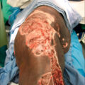
The clinical presentation of NEC can include abdominal pain and possible peritonitis, distension, feeding intolerance, and blood per rectum. Imaging may demonstrate dilated loops of bowel, pneumatosis intestinalis, portal venous gas, or pneumoperitoneum due to perforated viscus. A high index of suspicion with early diagnosis and intervention is vital for optimizing patient outcomes. New-onset abdominal pain, feeding intolerance, or burn sepsis of unknown etiology should raise concern for NEC, and broad-spectrum antibiotics should be considered. Patients who fail to respond to medical therapy and those who demonstrate peritonitis or pneumoperitoneum should be evaluated for surgical intervention. Frankly necrotic and/or perforated segments of bowel should be resected ( Fig. 31.4 ). In the event of questionable or patchy necrosis, particularly when extensive lengths of bowel are affected, the patient should undergo resuscitation with planned second-look operation within 24 to 48 hours to preserve as much bowel as possible and to reduce the risk of short gut syndrome.
Necrotizing enterocolitis.
( A ) Representative radiograph demonstrating multiple, dilated, persistently fixed bowel loops, an ominous sign for full-thickness necrosis of involved segments of bowel. ( B ) At operation, multiple necrotic bowel loops were found.
Clostridium difficile infection
Massively burned patients are at high risk for pseudomembranous colitis because they are frequently treated with multiple antibiotics for systemic infections. Clostridium difficile overgrowth can result in pseudomembranous colitis, with the potential to progress to fulminant toxic colitis and bowel perforation. Antibiotic stewardship is the key to preventing C. difficile infection (CDI). In a report of 180 thermally injured patients, the overall incidence of CDI was ∼8% with a mean burn TBSA of 42%. A potential complication of CDI is toxic megacolon, which increases patients’ risk of colonic perforation. Thus symptoms of colitis, such as leukocytosis, abdominal pain, distension, and grossly bloody diarrhea, must be promptly recognized. Stool samples are tested for C. difficile antigen. All unnecessary systemic antibiotic therapy should be discontinued, and appropriate CDI treatment should be initiated. The current Infectious Disease Society of America treatment guidelines recommend oral vancomycin or fidaxomicin for the first episode of CDI. For recurrent episodes, pulsed regimens of vancomycin or fidaxomicin are recommended. Fecal microbiota transplant should be considered in patients with multiple recurrent episodes. In patients with fulminant disease, which is characterized by the presence of megacolon, hypotension, or shock, vancomycin should be administered through a nasogastric tube in conjunction with intravenous metronidazole. Rectal vancomycin should be given to patients with concomitant ileus. Novel treatment strategies in development include the use of new antibiotics (ridinilazole, surotomycin, cadazolid), luminal antibodies (oral administration of whey protein concentrate prepared from hyperimmune bovine colostrum against C. difficile toxins), intravenous immunoglobulins, vaccines, and nontoxigenic C. difficile strains.
Surgical intervention may be required for patients with complicated or fulminant CDI. Operative intervention must also be considered in patients with progressive abdominal distension, peritonitis, shock, signs of sepsis, altered mental status, leukocytosis, lactic acidosis, or failure to improve after 5 days of medical therapy. Subtotal or total abdominal colectomy with end ileostomy has been advocated as the operation of choice. , An alternative surgical approach is to create a loop ileostomy with intraoperative colonic lavage using warmed polyethylene glycol 3350/balanced electrolyte solution (GoLYTELY) followed by postoperative vancomycin colonic flushes via the ileostomy in an antegrade fashion, with a reported mortality of 19% vs. 50% for patients undergoing colectomy. This operation can be performed laparoscopically with a high rate (80%) of reversal of ileostomy for GI tract continuity restoration. ,
Vascular complications
Suppurative thrombophlebitis
Suppurative thrombophlebitis is characterized by venous thrombosis associated with inflammation and bacteremia. Specific risk factors for suppurative thrombophlebitis include both burn injury and prolonged intravenous catheterization. The diagnosis may be made based on culture results together with evidence of thrombosis. The classic physical examination findings of edema, erythema, pain, and a palpable cord may not all be present. Burn patients frequently have positive blood cultures and clinical sepsis without an obvious source, and, in the setting of suppurative thrombophlebitis, the most commonly cultured organisms from infected veins reflect those cultured from the burn wound.
The incidence of suppurative thrombophlebitis complications in burn patients has been estimated to be as high as 7%, with significant risk in patients with greater than 20% TBSA burns, and it is associated with significant mortality. The principles of treatment include removing the source of infection, administration of intravenous antibiotics, and/or anticoagulation. Surgical treatment consists of surgical cutdown and evacuation of vein contents. If pus or clotting is found, the vein segment should be excised to a normal-appearing vein (usually up to the first uninvolved tributary). If exploration is negative at one site, then sequential exploration of other sites is necessary until the source of infection is identified. Prompt surgical intervention is necessary to prevent hematogenous dissemination and septic emboli that can result in endocarditis and osteomyelitis. The wound should be loosely packed and allowed to heal either by secondary intention or by delayed closure upon resolution of the infection.
To minimize the incidence of catheter-related complications, the current standard of practice for central venous access involves protocolized aseptic care, which includes full surgical attire with sterile technique and regularly scheduled catheter site dressing changes.
Central venous access
Adequate venous access is imperative in the acute postburn period for aggressive fluid resuscitation. Although large-bore peripheral intravenous catheters are the preferred route for resuscitating trauma patients, placement can be extremely difficult in those with major burns involving the extremities as well as peripheral vasoconstriction. Therefore central venous catheters have become standard practice in major burns. Particular attention should be given to the use of various available cannulation sites, depending on the size of the patient and considering the clinical condition of the burn wounds. The rate of catheter-related bloodstream infections in burn patients is estimated to be 20 per 1000 catheter days. Therefore compulsory site care is required. Routine central venous catheter exchanges in burn patients are controversial and typically performed based on institutional guidelines. When utilized, catheter replacement over a guidewire exchange is acceptable because it is more difficult to relocate lines in severely burned than in nonburned patients. However, catheters should not be exchanged over a guidewire if there is clinical evidence of infection, including erythema, drainage from the site, or bacteremia.
Central venous line placement is associated with potentially serious complications. To reduce the risk of complications, many burn centers rely on fluoroscopic or ultrasound guidance during the insertion of central venous catheters. Bleeding complications associated with central venous access vary in location and can be local, mediastinal, intrathoracic, or pericardial. Local hemorrhage typically occurs in patients with coagulopathy and can be controlled with local pressure. Hemorrhage into the thoracic space can occur at the time of catheter insertion, but it can also be seen when the catheter erodes through the vein wall. The most common situation leading to venous wall perforation occurs during the insertion of the percutaneous introducer sheath over a guidewire. As the sheath is introduced, it can fail to negotiate the path of the vein and traumatize the vein wall. Consequently, blood may accumulate into the mediastinum, pleural space, or pericardium. Small injuries typically resolve on their own. However, in cases of a larger venous injury, rapid bleeding into the thoracic cavity can occur, and emergency thoracotomy may be necessary. Bleeding or infusion of fluid into the pericardial space can rapidly compromise cardiac function and result in hemodynamic collapse. Pericardial tamponade typically presents with hypotension, muffled heart sounds, and distended neck veins (Beck triad). However, all components of the classic triad of symptoms are rarely present, and a physician must have a high index of suspicion to recognize this condition early. Echocardiography can confirm the clinical suspicion, and pericardiocentesis or pericardial window is therapeutic.
Related posts:


Stay updated, free articles. Join our Telegram channel

Full access? Get Clinical Tree


