Abstract
Deoxycholic acid (DCA) is an injectable adipocytolytic agent that is FDA-approved for the reduction of convexity or fullness associated with submental fat. When injected into the subcutaneous tissue, DCA induces adipocytolysis by irreversibly disrupting the cell membrane. Most standard markings for submental DCA injections target only a small central area of the submentum to avoid complications. This approach, however, often undertreats patients with adipose deposition outside the central region. The expanded safe zone (ESZ) system was developed based on cadaveric dye studies of submental fat compartments and describes a method for safely expanding the standard centralized treatment area. This technique provides topographical landmarks that correlate to discrete fat compartments within the preplatysmal fat and presents an anatomical basis for individualized treatment of submental convexity. By using the ESZ system to identify the zone of treatment, clinicians are able to: customize treatment strategy, improve jawline definition and cervicomental angle, and optimize the outcome of submental DCA injection.
93 Deoxycholic Acid Role in Fat Reduction
Key Points
Deoxycholic acid (DCA) is an injectable adipocytolytic agent approved for the reduction of convexity or fullness associated with submental fat.
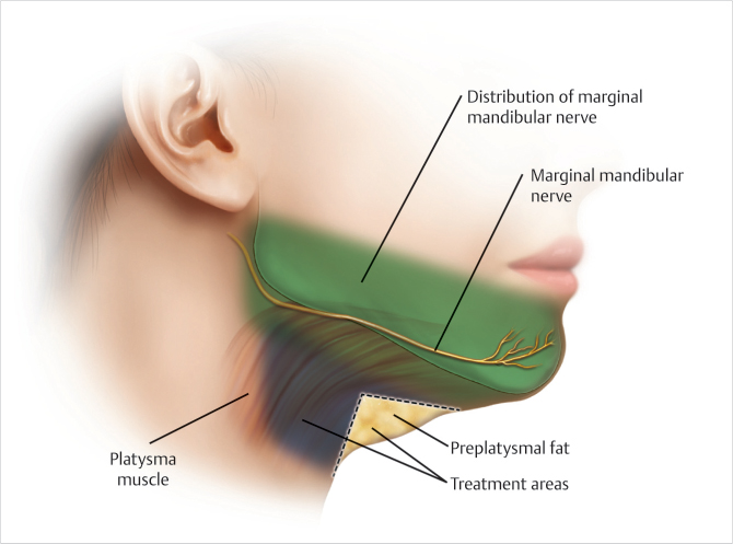
Most standard markings for DCA injections target a small central area of submental fullness to avoid complications, namely, marginal mandibular nerve paresis; this standardized treatment approach undertreats patients with adipose deposition outside the central region.
The expanded safe zone (ESZ) system describes a method for safely expanding the standard treatment region based on anatomical studies of submental fat compartments.
This technique provides topographical landmarks that correlate to discrete fat compartments within the preplatysmal fat and allows for targeted, patient-customized treatment of submental fullness.
93.1 Preoperative Steps
93.1.1 Analysis
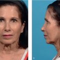
DCA injection to the submental region begins with a thorough analysis to assess the extent of fat deposition and identify the zone of treatment (Fig. 93.1).
Before proceeding with treatment, patients should be screened for other potential causes of submental fullness (e.g., low hyoid position, enlarged or ptotic submandibular glands, thyromegaly, and/or cervical lymphadenopathy).
93.1.2 Expanded Safe Zone (ESZ) System: Submental Markings
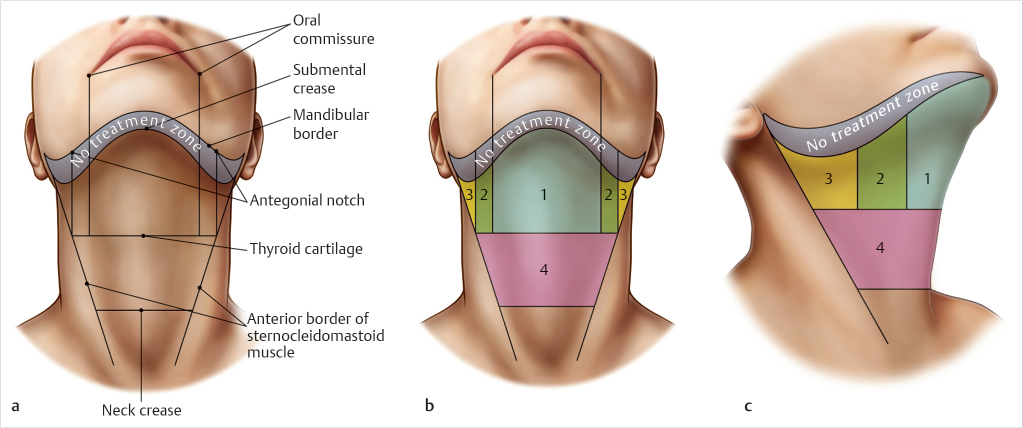
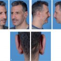
All six regions of the ESZ system should be assessed to identify the target treatment area.
The six regions of the ESZ system are marked (Fig. 93.2a) preoperatively in the seated and upright position based on the following anatomic boundaries:
No Treatment Zone (NTZ): Region 4.5 cm cephalad to the gonion and approximately 2 cm caudal to the inferior border of the mandible (corresponding to location of the marginal mandibular nerve) (Fig. 93.2b, c)
Safe Zone 1: Submental crease (superior border), thyroid cartilage (inferior border), caudal continuation of bilateral oral commissures (lateral borders) (Fig. 93.2b, c)
Safe Zone 2 (Bilateral): Inferior edge of the No Treatment Zone (superior border), lateral extension of thyroid cartilage (inferior border), caudal continuation of oral commissure (medial border), and antegonial notch (lateral border) (Fig. 93.2b, c)
Safe Zone 3 (Bilateral): Inferior edge of the No Treatment Zone (superior border), lateral extension of thyroid cartilage (inferior border), caudal continuation of antegonial notch (medial border), and anterior border of unilateral sternocleidomastoid muscle (lateral border) (Fig. 93.2b, c)
Safe Zone 4: Thyroid cartilage (superior border), neck crease (inferior border), anterior border of bilateral sternocleidomastoid muscles (lateral borders) (Fig. 93.2b, c)
93.1.3 Treatment Area and Injection Pattern
Outline the planned treatment area with a surgical pen, particularly avoiding the No Treatment Zone (Fig. 93.2b, c).
Planned treatment area is confirmed by palpation to ensure the presence of sufficient subcutaneous fat.
Prep treatment area with hypochlorous acid and apply a 1-cm injection grid to mark the injection sites.
Related posts:

Stay updated, free articles. Join our Telegram channel

Full access? Get Clinical Tree


