54 Distal Interphalangeal Joint Arthrodesis
Abstract
This chapter on distal interphalangeal joint (DIPJ) arthrodesis describes the techniques, indications, contraindications, and procedural pearls and pitfalls.
54.1 Description
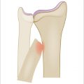
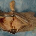
The distal interphalangeal joint (DIPJ) is a ginglymus (hinge) joint with an average arc of motion approximating 70 degrees. When diseased either as a result of trauma, rheumatologic conditions, or progressive deterioration of cartilaginous surfaces over time, the joint can become stiff, painful, and deformed. In these instances, eliminating the joint and repositioning the posture of the distal phalanx relative to the middle phalanx can provide pain relief and correction of deformity. This can be accomplished with a formal arthrodesis.
There are numerous methods of achieving a successful DIP fusion, all of which involve preparing the proximal and distal bone surfaces by removing remaining cartilage and cortical bone, positioning the fusion site to achieve optimal cancellous bone-to-bone contact, and utilizing a construct (pins, wires, screw, or combination thereof), to achieve skeletal stability.
54.2 Key Principles
Various methods of achieving skeletal stability exist. The selection of fixation device should take into consideration the bone stock, size of the medullary canal, and adequacy of the soft tissue envelope. (► Table 54.1)
Meticulous preparation of the bone is of paramount importance. The bone must be taken back to healthy appearing cancellous bone prior to fixation.
The germinal matrix must be protected during the fusion to avoid postoperative nail plate deformity.
The fusion is often positioned in neutral extension unless there exists a specific patient request or vocational need to have the digit flexed at this level.
54.3 Expectations
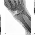
DIP fusions are well-tolerated, not technically demanding, have a favorable risk/benefit profile, and predictably provide patients with pain relief and satisfaction. The fusion construct requires a period of postoperative splinting until stability is either suspected clinically or confirmed radiographically. Postoperative imaging is not always necessary if the fusion site is clinically stable and patient free from pain. Postoperative therapy is rarely required unless the patient develops proximal interphalangeal joint stiffness or stiffness of adjacent digits. This fusion has a relatively low nonunion rate.
54.4 Indications
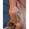
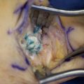
Disruption of a joint’s integrity brings with it the risk of limited motion, painful motion, or both. Deterioration of the distal interphalangeal joint can also be associated with clinical deformity (Heberden’s Nodes, angulation deformities, or mallet posture) and periarticular ganglion cysts (mucous cysts). Ganglion cysts at this level can cause attenuation of the overlying skin, become intermittently infected, and often result in nail plate deformity.
For the patients with limited motion and pain, or for those with gross clinical deformity, functional disturbance, or recurrent bothersome ganglion cysts, the physician should consider a motion eliminating treatment (arthrodesis). For most DIPJ derangements, a motion-eliminating treatment is predictable, well-tolerated, and carries a favorable risk-tobenefit profile.
54.5 Contraindications
Since a successful fusion requires a healthy and adequate bone stock as well as a suitable soft tissue envelope, contraindications to this procedure include active local skin infection, inadequate, scarred or noncompliant skin envelope, active bone infection, or inadequate bone stock. Further contraindications include unrealistic patient expectations (e.g., unwillingness to trade motion loss for pain relief) or a patient’s inability to comply with a postoperative splinting regimen.
For the patient with limited motion/deformity but no pain, no treatment is necessary unless the limited motion or deformity places the hand at a functional disadvantage.
Related posts:
Stay updated, free articles. Join our Telegram channel

Full access? Get Clinical Tree


