28 Anterior Trunk Disorders
Summary
Disorders of the anterior trunk include congenital defects of the abdominal or chest wall. Gastroschisis and omphalocele are the two most common congenital abdominal wall defects. The absence of a covering membrane around the herniated abdominal contents differentiates gastroschisis from omphalocele, and also may affect timing of surgical management. Bladder exstrophy is a rare malformation of the infra-umbilical abdominal wall that is easily recognizable at birth, but is rarely diagnosed prenatally. Prune belly syndrome is characterized by hypoplastic or absent anterior abdominal wall musculature leading to a wrinkled appearance of the abdomen. Congenital chest wall diagnoses include pectus excavatum and pectus carinatum. Pectus excavatum is characterized by a concave appearance to the anterior chest wall, while pectus carinatum presents as a protrusion of the sternum and/or ribs with a depression along the sides of the chest.
The goal of surgery for patients with gastroschisis and omphalocele is to safely return the eviscerated bowel to the peritoneal cavity and establish abdominal wall integrity. Surgical closure for gastroschisis and omphalocele can be achieved via primary reduction, or delayed, staged closure. Treatment for bladder exstrophy usually occurs in a staged manner to close the bladder and abdominal wall. Plastic surgeons may be involved in late corrections of abdominal scarring and, incisional hernias or bulges. Although the majority of umbilical hernias will close spontaneously, large defects or those that persist to school age may require surgical repair. A variety of non-operative, minimally invasive and invasive modalities exist to treat both pectus carinatum and excavatum.
28.1 Introduction
Disorders of the anterior trunk constitute a wide array of congenital defects of the abdominal or chest wall. These disorders range in severity and frequency and are typically managed by pediatric surgeons. Plastic surgeons who treat children and young adults can apply a host of reconstructive and aesthetic techniques in the early as well as late management of these conditions. As such, it is worthwhile for the plastic surgeon to familiarize themselves with some of the more common anterior trunk disorders.
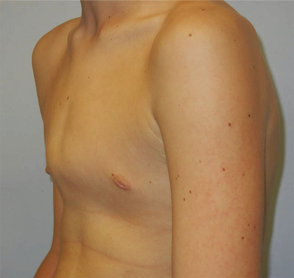
Minor congenital abdominal wall defects are common. In particular, umbilical or inguinal hernias are frequently encountered. Gastroschisis and omphalocele are the most common serious congenital abdominal wall defects, and are frequently diagnosed antenatally. Gastroschisis is characterized by free herniation of the abdominal viscera. There is no membrane covering the herniated abdominal contents, which differentiates gastroschisis from omphalocele. This condition has doubled in incidence over the past decade for unclear reasons, with approximately 2,000 new cases per year in the United States. Omphalocele is a related condition also characterized by evisceration of intra-abdominal organs. However, these viscera are contained in a sac that protects the viscera, minimizes neonatal fluid losses, and affords substantial time to repair unlike gastroschisis (Fig. 28‑1). Unlike the periumbilical defects above, bladder exstrophy is a rare malformation of the infraumbilical abdominal wall. Incomplete closure of the bladder, protrusion of the posterior bladder wall through the lower abdominal wall, epispadias, as well as alterations in the pelvic bones and muscles necessitate early acute multidisciplinary care. Late defects of the abdominal wall such as bulges, hernias, and depressed scars are common. In addition, complex genitourinary defects may also require multiple complex reconstructive procedures. Another very rare condition that affects both the abdominal wall and the urinary tract is prune belly syndrome. In this condition, the anterior abdominal wall musculature is hypoplastic or absent, leading to a wrinkled appearance of the abdomen (Fig. 28‑2). In addition to the marked skin redundancy and distortion of the trunk, severe urological abnormalities and cryptorchidism frequently occur.


The range of a congenital chest wall diagnoses is smaller, but some diagnoses are common as well. Pectus excavatum, for example, may occur in 1/150 to 1/1,000 live births and is familial in 40% of cases. It is characterized by a concave appearance to the anterior chest wall and is the most common chest wall abnormality. The depression may be mild or quite severe, impacting cardiac and pulmonary function (Fig. 28‑3). In addition, it can be present at birth or manifest during adolescence. In contrast, pectus carinatum presents as a protrusion of the sternum and/or ribs with a depression along the sides of the chest (Fig. 28‑4). Though not as common as pectus excavatum (occurring only 1/10th as often), it accounts for roughly 5 to 7% of chest wall anomalies overall. The condition is usually isolated and nonsyndromic though pectus carinatum can be seen in patients with Marfan, Ehlers–Danlos, Noonan, and Turner syndromes as well as others.

28.2 Diagnosis
Most diagnoses of the anterior trunk can be made following history and physical examination. Radiological evaluation may also be extremely useful early or even later in life. Severe conditions can also be recognized by prenatal ultrasound and appropriate plans for postdelivery management made in advance. In many of these conditions, concurrent severe comorbidities may exist and must be sought out.
The prenatal diagnosis of gastroschisis is possible from the end of the first trimester, after the physiological closure of the abdominal wall around 10 weeks of gestation. The ultrasound detects loops of bowel outside of the abdominal cavity, herniated through a small paraumbilical wall defect and floating in the amniotic fluid without any covering membrane. The absence of this covering allows one to differentiate gastroschisis from omphalocele. Present techniques can predict complicated gastroschisis and outcomes associated with degree of intra-abdominal dilatation. A raised maternal alpha fetoprotein is also indicative of gastroschisis. Gastroschisis is not associated with chromosomal anomalies and generally occurs as an isolated anomaly. An omphalocele can also be diagnosed by ultrasound during the late first trimester, but is more commonly confirmed during the 18-week ultrasound. In some cases, an elevated maternal alpha fetoprotein level may also be identified, though less commonly than in gastroschisis. Associated anomalies are common and include cardiac, renal, skeletal, and neural tube abnormalities. Omphaloceles are often associated with chromosomal anomalies (such as Trisomy 13, 18, and 21) as well as other syndromes such as Beckwith–Wiedemann.
Umbilical hernias occur when the umbilical ring is not fully closed and can be diagnosed upon physical examination. Rarely is any imaging required. A protruding soft-tissue mass and a palpable defect in the fascia is diagnostic. Similarly, pediatric inguinal hernias are diagnosed clinically with a visible soft-tissue bulge in the inguinal region. In boys, ipsilateral scrotal swelling may be secondary to a hernia or a hydrocele. Ultrasonography may be used to differentiate between the two.
Bladder exstrophy is easily recognizable at birth, but is rarely diagnosed prenatally. The prenatal diagnosis consists of a nonvisible fetal bladder and low insertion of the umbilical cord. Prune belly syndrome can be diagnosed through a prenatal ultrasound, although diagnosis is usually made shortly after birth. It occurs most often in male infants. The ultrasound diagnosis in early gestation requires the identification of a megacyst (distended bladder) and oligohydramnios. Undescended testes and urinary tract anomalies are often noted postnatally on physical examination and imaging.
The pectus excavatum and carinatum deformities are easily visualized on physical examination. In some, the deformity may be present early in life, whereas in others it will manifest during adolescent growth. Symptoms may include exercise intolerance, decreased endurance, pain, body image issues, and dyspnea. In symptomatic individuals, computed tomography (CT) scans may be warranted to determine the severity of the pectus defect and potentially assist in reconstructive planning. In cases of pectus excavatum, cardiac and pulmonary compression may be documented in conjunction with functional tests such as an echocardiogram or pulmonary function testing. Patients with pectus carinatum manifest a sternal protrusion with a depression along the sides of the chest. Most are male and present during adolescence. Associated symptoms including chest wall pain, frequent injuries, body image issues, and scoliosis should be sought. Displacement of the thoracic musculature can interfere with breathing, and 10% of patients experience exercise intolerance. In symptomatic patients, a chest CT scan in conjunction with pulmonary function testing can document severity.
Related posts:


Stay updated, free articles. Join our Telegram channel

Full access? Get Clinical Tree


