27 Perforator Flap Reconstruction of Breast-Conserving Therapy Deformities
The treatment of early stage breast cancer by partial mastectomy followed by postoperative radiotherapy, often referred to as breast conservation therapy (BCT), may be considered as an oncologic equivalent to mastectomy in selected cases. 1 , 2
After partial mastectomies (lumpectomies and quadrantectomies) that preserve the nipple-areola complex (NAC) and a large portion of the native breast tissue, up to 31% of the patients indicate an unsatisfactory appearance after BCT. 3 – 5 Most conditions that lead to the described poor aesthetic outcome following BCT are a result of the surgical dilemma in the treatment of breast cancer. This dilemma arises because wider excision is necessary to provide clear margins and better local control of breast cancer on the one hand, and because of the concerns about sparing as much tissue as possible for defect closure and the resulting aesthetic outcome on the other hand. 6 – 8
Our preference is to perform immediate reconstruction when it is feasible and indicated. To obtain a satisfactory aesthetic result, the created cavity should be filled with local or distant tissues before starting the irradiation, because operating on irradiated breasts has high complication rates and often poor aesthetic results. During immediate reconstruction, the breast can be manipulated before irradiation, this could lower the complication rate and improve the outcome. 7 – 10
The clinical outcomes and expert impressions support our approach. 8 – 15 They suggest that surgical factors are more important than radiation-induced injury and fibrosis in the development of post-BCT breast deformity. When immediate reconstruction is not performed in cases with unfavorable resection defects, significant breast deformities will likely manifest following completion of the BCT regimen. There are many reasons for aesthetic failure:
Tumor resection can produce distortion, retraction, or volume changes in the breast.
Changes in the nipple-areola complex can exaggerate asymmetry.
Irradiation can have a profound effect on the remaining breast tissue; however, it is very difficult to predict who will develop those severe postradiation changes. 7 , 16 Radiotherapy generally “exaggerates” the surgically created deformity. 17
Several classification scales have been developed to characterize delayed breast deformities, and they suggest reconstructive options. Berrino et al 15 were the first to classify post-BCT deformities. Their morphologic classification system underscores the importance of analyzing the etiologic factors of the deformity. 15 , 16 They described the following four deformity types, which are based on the presence of one or more: nipple malposition, localized breast defect, generalized breast contracture, or severely total damaged breast. 15
Clough et al 13 have altered this classification, based on response to reconstruction.
In clinical application, these classifications help to clarify the deformities, which typically result when BCT is performed under suboptimal conditions. The classification schema also guides us in reconstruction by emphasis on identifying what is missing or disordered and on seeking a reasonable match between the two breasts. In this chapter, we will focus on post-BCT deformity which required surgical procedures (mainly flap surgery) to the irradiated breast.
Clinical Approach to BCT Deformities
As for all reconstructive procedures, reoperative breast surgery should be carefully planned on an individual base. Before considering any treatment, relapse of cancer should be ruled out.
In the approach to BCT deformities two major points should be considered:
Fewer local options of repair methods and techniques are available in breasts previously treated with BCT because of reduced breast volume, scarring, distorted anatomy, and disturbed vascularity.
Postradiation changes make corrections more technically difficult, and results and complications are highly unpredictable.
Therefore an extensive surgical procedure of the breast after BCT should be carefully planned, because of the high complication rate, including wound dehiscence, fat necrosis, skin necrosis, and nipple necrosis. 8 , 13 Studies have estimated the complication rate to be as high as 50% 7 and the final aesthetic result to be poor when extensive tissue rearrangement is performed in a previously irradiated breast. 13 , 18
Preoperative Analysis of the Deformity
Delayed partial breast reconstruction should be considered when post-BCT deformities occur. However, the patient’s expectations for the cosmetic outcome are often much higher than during the primary cancer treatment phase. Therefore thorough preoperative analysis of the problem and the patient’s expectations should be explored during several outpatient visits. Careful surgical planning and clear patient consent is essential, as in every reoperative surgery.
The clinical examination should encompass the patient’s general condition and any risk factors such as obesity, smoking history, or any concurrent disease, such as diabetes mellitus or arterial hypertension. A high BMI and smoking history are major risk factors that adversely affect wound healing, compounding the vulnerability of the tissue as a result of irradiation. Patients should be instructed to quit smoking at least 3 months before surgery.
A thorough oncologic checkup is essential before planning a surgical procedure in patients after BCT deformity. Because these patients have a greater risk of local recurrence than mastectomy patients do, clear documentation of initial tumor therapy and margin status is essential.
Local physical examination includes both breasts and the surrounding tissue, such as the axillary region, shoulder, and back. Any limitation of the ipsilateral breast function should be documented. Although correction of irradiated breast tissue by removing scar tissue and adding “healthy” flap tissue may improve the locoregional aspect and function, functional deterioration may result if a major flap-related complication occurs.
When the irradiated breast is analyzed, several points should be considered:
Breast volume and position
NAC position
The number of affected quadrants
The general quality of the breast
Depending on the resultant deformity, one or more surgical options can be performed. Regardless of the reconstructive method that is required for the irradiated breast, a contralateral breast remodeling may still be necessary to achieve better breast symmetry. Therefore the contralateral breast is almost a part of the surgical strategy and should be discussed with the patient.
Reconstructive Methods for BCT Deformities
The ultimate goal of the treatment is to achieve breast symmetry. We consider the treatments of BCT deformities to be salvage procedures. The currently available methods in reconstructive surgery cannot re-create the initial anatomy status of the breast, which is irreversibly damaged by previous surgery and irradiation. This last point must be emphasized to the patient during the preoperative visit.
The available surgical methods for BCT deformities are divided into three groups. Nevertheless, the definitive surgical plan may still include more than one option.
Techniques That improve Breast volume and Position
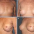
When there is no major deformity in the irradiated breast, procedures on the contralateral breast to adjust symmetry are ideal options. However, BCT often results in a smaller and higher position of the breast. In these cases, either a breast fat grafting procedure 19 or contralateral breast remodeling or a combination of both is the optimal treatment. The contralateral breast is often larger and more ptotic.
Techniques That improve NAC Position
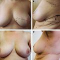
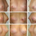
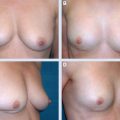
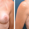
The techniques described may be adequate to correct the asymmetry in the NAC position. However, when a horizontal displacement of the NAC is found, additional repositioning is required. Mild lateral or medial NAC shift can be corrected with elliptic deepithelialization or with a circumferential or round bloc mastopexy. 14 When mild radiation sequelae are observed, a bilateral matching procedure can be done. 8 , 12 , 16 Nevertheless, techniques in mammaplasty should be adapted to this specific situation. A minimal skin undermining with a short and wide pedicle must be used. 8 When a major NAC malposition is caused by a large breast defect or a generalized severe irradiation sequela, flap surgery is indicated.
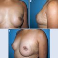
Techniques That Correct Breast Defects
Box 27-1 General Guidelines for Perforator Flap Reconstruction After BCT
Excision lines have to be designed within the reconstructive approach.
The breast tissue should be opened like scar release after burn contracture.
Fibrosis and scar tissue is removed without undermining of the breast tissue.
Frozen section examination can be requested if recurrence is suspected within the excised tissue.
Flaps should only be incised after releasing the breast for better evaluation of the breast defect.
Related posts:
 28 Local Perforator Flaps in Oncoplastic Breast-Conserving Surgery: The Nottingham Experience
 26 Correction of the Breast-Conserving Therapy Deformity Using Local Flaps
 29 Breast Lumpectomy Reconstruction With External Vacuum Expansion and Autologous Fat Grafting
 31 Can Implants Correct a Breast-Conserving Therapy Deformity?
 30 Fat Grafting in the Breast-Conserving Therapy Deformity
 24 Classification and Analysis of the Breast-Conserving Therapy Deformity
Stay updated, free articles. Join our Telegram channel

Full access? Get Clinical Tree


