10 Bipedicled (Double or Stacked) Abdominal Perforator Flap in Unilateral Breast Reconstruction
Julie V. Vasile and Joshua L. Levine
The abdomen remains an ideal choice of donor site for autologous breast reconstruction. In unilateral breast reconstruction, sometimes one hemiabdominal flap, based on the ipsilateral deep inferior epigastric perforator (DIEP) pedicle, does not provide enough volume for a reconstruction that matches the contralateral breast. Adding the contralateral hemiabdomen with its own perforator flap pedicle is an excellent method to increase the volume of perfused fat and skin. The larger, bipedicled flap allows for much greater control in achieving symmetry and ptosis. This surgery is particularly helpful in the radiated or delayed breast reconstruction with a significant skin deficit compared with the contralateral breast. It can also improve success when trying to match a large ptotic breast.
Adding the second hemiabdomen should not be thought of as simply performing another abdominal perforator flap. The addition of the second flap adds to the complexity of the reconstruction for a variety of reasons. Although two flaps are harvested, a midline abdominal incision is not usually made.1,2 Therefore, the options for approaching the perforators are limited. The second pedicle also has implications with respect to recipient vessel selection and dissection, and if a flap-to-flap anastomoses is to be considered, a suitable branch point off of the primary pedicle must be considered. The increased complexity of the microsurgery influences how the flaps are inset, and postoperative monitoring must be adjusted to take dual perfusion into account. The increased complexity of bipedicled DIEP flap procedures makes them more challenging than a unilateral abdominal perforator flap, but with proper planning, obstacles can be anticipated and overcome, and the bipedicled abdominal perforator flap can be a powerful and rewarding operation for breast reconstruction.
Indications
The bipedicled abdominal perforator flap is considered for unilateral breast reconstruction, especially in thin patients who need a substantial amount of volume to match the contralateral breast. Prior abdominal surgery is not an absolute contraindication, provided that there is enough tissue to reconstruct a breast.
Imaging for Bipedicled Abdominal Perforator Flaps
Preoperative magnetic resonance angiography (MRA) evaluation of the vessels is important for maximizing success and decreasing anesthesia time. The first step is to determine which vessels perfusing each hemiabdomen are best. Most commonly, the DIEP is considered the best vessel because of its larger size and perfusion of thicker fat located along the central lower abdomen. Alternative vessels could be the superficial inferior epigastric pedicle or superficial circumflex iliac pedicle. The second step is to try to determine the ease of connecting one hemiabdominal flap pedicle to the other, and at which locations along the pedicles this may be possible. Each DIEP pedicle is evaluated with respect to its branching pattern. Large branches are compared with the size of the vessels at the cephalad continuation of the pedicle beyond the perforator. If either of these vessel options is robust, flap-to-flap perfusion should be considered. The superficial inferior epigastric arteries are also evaluated.
When evaluating the vascular anatomy, the approach to the dissection should also be considered. If the best perforators are in the medial row of either flap, the microsursdgeon must be ready to anticipate the sacrifice of the lateral row, because the medial vessels cannot be viewed via a midline incision.
Donor-Site Dissection
The abdomen is not bisected along the midline. Both hemidabdominal flaps can still be dissected simultaneously, with each surgeon starting laterally or moving from cephalad to caudal. When the lateral perforators are encountered, a vessel is either selected or sacrificed with the knowledge that a better vessel exists further medial (as seen on MRA). Sometimes lateral perforator options can be preserved while confirming the larger perforators medially. Alternatively, one can learn to trust the MRA image and proceed directly to the perforator of choice, or if one DIEP pedicle dissection is completed first, the pedicle can be transected, and dissection can continue across the midline to approach the second DIEP pedicle medially.
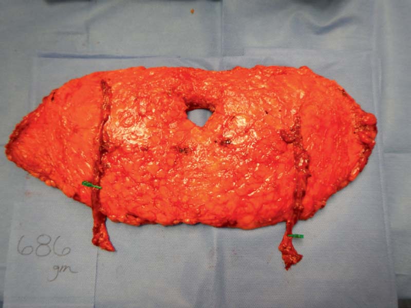
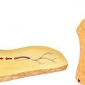
The DIEP pedicle is dissected in the typical manner except for two important points. First, the cephalad continuation and DIEP pedicle branch are dissected for a length adequate for anastomoses if the plan is to connect one pedicle to the other. Second, the DIEP pedicle is dissected caudally until near the external iliac vessels to yield a long pedicle. Dissecting a long pedicle (>8 cm) will aid in decreasing tension on the vessels during microsurgical anastomosis and insetting ( Fig. 10.1 ).
Related posts:
 2 Starting a Perforator Flap Breast Program
 4 Deep Inferior Epigastric Perforator Flap for Breast Reconstruction
 9 Pedicled Lateral Thoracic Flap for Breast Reconstruction
 13 Lateral Thigh Perforator Flap (Septocutaneous Tensor Fasciae Latae Perforator Flap)
 15 Deep Circumflex Iliac Artery Perforator Flap for Breast Reconstruction
 11 Venous Salvage Procedures in the Deep Inferior Epigastric Perforator Flap for Breast Reconstruction
Stay updated, free articles. Join our Telegram channel

Full access? Get Clinical Tree


