von Langenbeck Palatoplasty
Kathryn V. Isaac
David K. Chong
DEFINITION
Cleft palate is the failure of fusion of the palatal shelves during embryological development.
There are many surgical techniques used for cleft palate repair. The repair technique chosen varies depending on the type of cleft palate and beliefs regarding facial growth and speech repercussions1.
ANATOMY
Embryologically, the cleft may affect the primary or/and secondary palate.
Anatomically, the cleft is defined according to the involvement of the hard and soft palates.
A complete cleft of the palate is defined as a cleft involving both the primary and secondary palates. Hence, a complete cleft involves the soft palate and the hard palate.
The Veau classification (FIG 1) provides a clinically useful description of the cleft type according to the involvement of the alveolus, the hard palate, and the soft palate. It guides selection of the surgical treatment.2
The von Langenbeck palatoplasty is most commonly employed for incomplete cleft palates, where the primary palate is unaffected (ie, soft palate and hard palate to incisive foramen) or where the primary palate was previously repaired (commonly with a superiorly based vomerine flap at the time of lip repair).1,3
Von Langenbeck palatoplasty is less commonly used to repair isolated clefts of the soft palate (Veau I) and mostly employed to repair clefts of the soft palate and secondary palate [hard palate posterior to the incisive foramen (Veau II)].
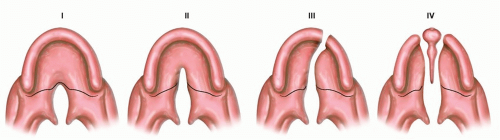
FIG 1 • Veau classification of cleft palate types. I. Cleft of soft palate only. II. Cleft of secondary hard and soft palates. III. Unilateral cleft extending through secondary hard and soft palate, through right or left primary palate, and through the alveolar process. IV. Bilateral cleft through secondary hard and soft palate, through bilateral primary palate and bilateral alveolar processes.
This technique may also be modified for complete clefts and is named a hybrid procedure.4 This hybrid procedure employs the bipedicled von Langenbeck flap on the greater segment and a unipedicled flap on the lesser segment. It is not used for the treatment of submucous clefts.
Additionally, the von Langenbeck technique of a bipedicled mucoperiosteal flap may be used for the repair of fistulae located in the hard palate or junction with the soft palate.
PATHOGENESIS
The etiology of cleft palate is multifactorial: environment, teratogen exposure (alcohol, anticonvulsants, steroids, diazepam), nutrition, maternal infections (rubella, toxoplasmosis), and genetics.
Cleft palate repair restores anatomical closure between the oral and nasal cavities.
An unrepaired cleft palate may result in impaired breathing, feeding, speech, and hearing.
NATURAL HISTORY
Orofacial clefting is the most common craniofacial birth defect, occurring in 1:750 live births.5
The incidence of cleft palate alone is 1:2000, with a female-to-male ratio of 2:1.
The incidence of cleft palate in combination with cleft lip is greater, estimated at 1:1000 with a reverse gender ratio.
In the population of children with cleft lip and/or cleft palate (CL+/-P), approximately 50% will have CLP, 30% to 35% CP, and 15% to 20% CL.
Approximately 40% of CP patients will have a syndrome, and 15% of CLP will have a syndrome.
PATIENT HISTORY AND PHYSICAL FINDINGS
The diagnosis of a cleft palate is made by prenatal ultrasonography or by physical examination at birth and in early infancy. A detailed assessment is made of the child’s breathing, feeding, growth, and development.
Airway assessment must exclude the presence of sleep apnea and micro-/retrognathia.
Assessment of growth and weight is key to identify and monitor feeding difficulty, gastroesophageal reflux, and the use of feeding aids (Haberman/Pigeon teat).
Hearing and middle ear function must be assessed and treated early by an otolaryngologist.
The plastic surgeon should define the cleft palate type according to the anatomic structures involved, the laterality, width of the palate, and the presence/absence of a cleft lip.
Also, the child should be assessed for associated anomalies suggestive of a syndrome: craniofacial dysmorphism, airway compromise, cardiac defects, ocular and auricular abnormalities, and musculoskeletal anomalies. A genetics referral is suggested if suspicious for a case of syndromic cleft palate.
DIFFERENTIAL DIAGNOSIS
Syndromic cleft palate
Van der Woude syndrome
Stickler syndrome
Robin sequence
22q11.2 deletion syndrome
Treacher Collins syndrome
Nager syndrome
Ectrodactyly-ectodermal dysplasia
Klippel-Feil syndrome
NONOPERATIVE MANAGEMENT
It is very uncommon not to repair a cleft palate.
If the child is unable to undergo surgical treatment secondary to airway concerns or medical fitness, a palatal obturator may be considered.
Such patients should be reassessed on a regular basis (at least annually), as they may become sufficiently stable for delayed surgical correction of the cleft palate.
SURGICAL MANAGEMENT
Cleft palate repair is ideally performed between 9 and 12 months of age, with the infant weighing greater than 8 kg.
Airway and middle ear disease assessment is performed and treated prior to or simultaneously with cleft palate repair.
Risks of the procedure are hemorrhage/hematoma, airway compromise, wound dehiscence, infection, oronasal fistula, flap necrosis, velopharyngeal insufficiency, and reduced midfacial growth.
The main objectives of the cleft palate repair are:
To separate the oral and nasal cavities
To reposition to the velar musculature for restoration of the velopharyngeal sphincter, in particular the reconstruction of the levator muscular sling
To improve Eustachian tube function
To minimize detrimental effects on subsequent maxillary growth
The goals of the palatoplasty are to improve feeding and speech.
The main steps of the procedure are:
Marking and elevation of bipedicled mucoperiosteal flaps off the hard palate
Dissection and separation of the nasal mucosa, velar musculature, and oral mucosa
Careful dissection of pedicles (greater palatine artery)
Tension-free closure of nasal mucosal layer
Reconstruction of the velopharyngeal sphincter with repositioning of the levator sling
Recreation of the uvula
Closure of the oral mucosal layer
Preoperative Planning
With the patient anesthetized, the width of the cleft is measured at the junction of soft and hard palate. The width of the cleft is important to predict difficulty of the cleft palate repair and anticipated amount of dissection required for closure.
The von Langenbeck technique provides less flap mobilization relative to other techniques (two-flap palatoplasty). Thus, this technique is best reserved for narrow clefts.
Positioning
The patient is positioned supine with shoulder roll to allow neck extension. An oral Rae tube, with the circuit directed caudally, is carefully taped centrally. Corneas are protected with eye tapping.
Access and visualization of the entire palate are very important and cannot be understated. It is especially important to ensure visualization from the uvulae to the anterior extent of the palate.
It is essential for the surgeon to ensure positioning is correct prior to scrubbing. This will facilitate the procedure.
The surgeon should have a headlight or lighted instruments for optimizing visualization in the confined space and a skilled attentive assistant.
It is advantageous to have a bed that has Trendelenburg positioning capability. Some surgeons stand to perform palate surgery from below, but having the baby’s head supported with the surgeon seated operating upside down provides the best exposure and is the preferred position by most surgeons.
Approach
The aim of cleft palate surgery is tension-free closure of the palate oral and nasal mucosal layers and restoration of the velopharyngeal muscular sphincter.
Difficulties anticipated include:
Access
Lighting
Bleeding obscuring visualization
Surgical approaches differ with regard to the amount of dissection performed to achieve tension-free closure and muscle repositioning.
Critical step is adequate exposure and dissection of tissues to provide comfort for the operating surgeon to achieve tension-free closure.
Oral layer mobilization is largely dependent on careful mobilization of the greater palatine artery from its periosteal sleeve.Related posts:

Stay updated, free articles. Join our Telegram channel

Full access? Get Clinical Tree