Vitiligo
Jeffrey T.S. Hsu
I. BACKGROUND
Vitiligo is a common acquired pigmentary disease affecting 1% to 2% of the population. Localized or generalized areas of the skin become completely depigmentated as melanocytes are selectively destroyed. This finding is in contrast to albinism, in which melanocytes are present but there is little or no pigmentation because of faulty or absent melanin synthesis. The exact cause of vitiligo remains unknown. Because of the association with other autoimmune diseases, an autoimmune etiology is favored.
Vitiligo appears to have a familial incidence of 20% to 30% and is found with increased frequency in patients with endocrinopathies. These include thyroid disease such as Hashimoto thyroiditis and Graves disease, other endocrinopathies such as Addison disease, gonadal atresia, and diabetes mellitus. In patients with vitiligo, there is also an increased incidence of halo nevi, pernicious anemia, alopecia areata, and ocular abnormalities. Complete spontaneous repigmentation is rare.
II. CLINICAL PRESENTATION
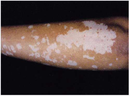
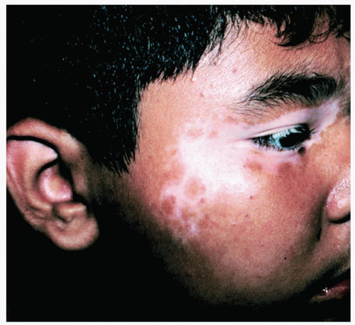
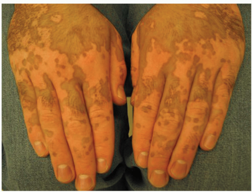
Onset of vitiligo is typically seen in ages 10 to 30, and rarely in infancy or old age. The lesions of vitiligo appear as white or depigmentated macules or patches, usually with sharp borders and are found symmetrically over boney prominence such as the hands, forearms, feet, and around body orifices (lips, eyes, and anogenital areas) (Figs. 46-1, 46-2, and 46-3). Lesions begin ranging from millimeters in size but can enlarge centrifugally at an unpredictable rate. Injury to the skin of these patients may cause a temporary or permanent loss of pigment in that area (Koebner phenomenon). Scars, scratch marks, and bruises may therefore heal with no pigment present. Hair growing from vitiliginous skin may be depigmented. There are no symptoms associated with the depigmentation of vitiligo. These areas, however, sunburn easily.
There are three subtypes of vitiligo: localized, generalized, and universal. Localized vitiligo involves less than 20% of the body surface area. It can be either focal, presenting as one of more macules in one area, most commonly in the distribution of the trigeminal nerve; or segmental, presenting in a dermatomal pattern. Segmental vitiligo is much more common in children than adults and tends to spread rapidly within the segment of skin (Fig. 46-4). This subtype is rarely associated with autoimmune or endocrine disorders. Generalized vitiligo can be found in more than one isolated area, usually acrofacial (involving distal fingers and around orifices) and/or widely scattered throughout the body. Universal vitiligo describes complete or near-complete depigmentation (Fig. 46-5). It is often associated with endocrinopathies.
III. WORKUP
The diagnosis of vitiligo is usually made with clinical observation (Table 46-1). A Wood lamp examination may be helpful to accentuate the vitiligo patches from normal skin, especially in fair skin. At times biopsies
may be required to rule out other causes of depigmentation and hypopigmentation. Histology shows complete absence of melanocytes and epidermal pigmentation.
may be required to rule out other causes of depigmentation and hypopigmentation. Histology shows complete absence of melanocytes and epidermal pigmentation.
TABLE 46-1 Differential Diagnosis | |
|---|---|
|
 Figure 46-2. Vitiligo on the forearm. Neutrogena Skin Care Institute. (Sauer GC, Hall JC. Manual of Skin Diseases. 7th ed. Philadelphia, PA: Lippincott-Raven; 1996.) |
Stay updated, free articles. Join our Telegram channel

Full access? Get Clinical Tree


