Introduction
In this chapter, the venous system of the head and neck is categorized into the veins of the face, scalp, and neck. The main venous drainage pathway of the face is through the hemiloop-like vein that surrounds the orbit. The vein can be formed by the supraorbital, angular, or facial veins, depending on its location. It collects most of the blood from the face and connects mainly to the zygomatico-temporal, superior ophthalmic, deep facial, and internal jugular veins. The main venous drainage pathway of the superficial parts of the scalp is through the superficial temporal, middle temporal, occipital, and posterior auricular veins. These veins drain into the external jugular vein. The internal jugular vein is the main venous drainage pathway of the head and neck; the external and anterior jugular veins are the pathways from the superficial layers of the region. The vertebral vein collects blood from the prevertebral muscles and drains into the brachiocephalic vein.
Veins of the Face
The main venous drainage pathway of the face is primarily the facial vein. In the middle of the face, the hemiloop-like vein surrounds the orbit.1 This vein can be contributed to by the supraorbital, angular, or facial veins, depending on its location (Fig. 7.1). These veins are connected to the zygomatico-temporal vein in the upper lateral region of the orbit, to the superior ophthalmic vein in the medial canthal area, to the deep facial vein in the nasolabial area, and to the internal or external jugular veins in the lower lateral area (Fig. 7.2). As with most superficial veins, these veins have many variations. Common patterns are discussed here (Fig. 7.3).
Supraorbital Vein
The supraorbital vein passes medially above the orbital rim under the orbicularis oculi muscle to connect to the angular vein in the medial canthal area. A branch of the supraorbital vein also connects to the superior ophthalmic vein at the supraorbital notch or foramen. Laterally, it connects to the zygomatico-temporal vein arising from the middle temporal vein near the zygomatic process of the frontal bone. There, it also connects with radicles of the superficial temporal veins.
Surgical Annotations
The zygomatico-temporal vein is known as a sentinel vein. The vein is located in a 10-mm zone above which the temporal branch of the facial nerve passes.2
Supratrochlear Vein
Basically, one or two large veins arise from the medial canthal area and run toward the forehead. The supratrochlear vein connects to the tributaries of the superficial temporal veins to form a large venous network in the forehead. Both the deep veins from the pericranial layer and the superficial veins from the galea frontalis layer empty into the vein.3 The supratrochlear vein finally joins the angular or nasal root vein near the medial canthus.1
Nasal Root Vein
Arising superficially from the angular vein, the nasal root vein pierces the procerus muscle and anastomoses with its contralateral counterpart to form a large communicating vein under the skin of the nasal root.1 The nasal root vein is convex toward the nasal tip. It is a bridge of the bilateral hemiloop-like veins. Several tributaries from the external nose connect to the nasal root vein (Fig. 7.4).
Angular Vein
The angular vein is formed by the union of the supratrochlear and supraorbital veins. It runs inferiorly across the medial margin of the medial canthal tendon approximately 8 mm from the medial canthus of the eye.4 It becomes the facial vein at its junction with the superior labial vein.5 The two major veins arise from it, namely, the transverse nasal root vein (superficial) and a branch to form the inferior root of the superior ophthalmic vein (deep). Several tributaries from the external nose and lower eyelid also connect to the angular vein (Fig. 7.5).
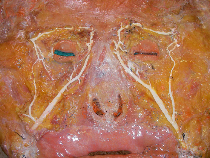
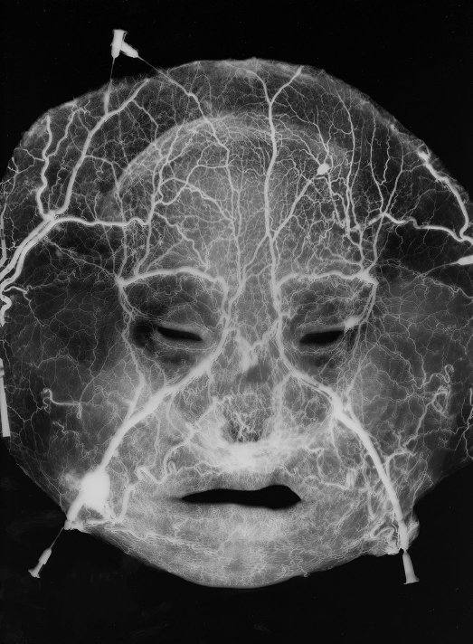
Fig. 7.2 Arteriovenogram of the face. The hemiloop-like vein surrounds the orbit. The tributaries of the superficial temporal vein connect to the vein in the forehead area. The supratrochlear vein connects to the vein in the medial canthal area. The vein drains into the facial vein.
Superior Ophthalmic Vein
The superior ophthalmic vein is formed at the superomedial margin of the orbit immediately posterior to the trochlea by the union of two contributing roots, namely, a superior root from the supraorbital vein and an inferior root from a branch of the angular vein. It runs with the ophthalmic artery and links the facial and intracranial veins. It traverses the superior orbital fissure to end in the cavernous sinus. The superior ophthalmic vein has venous valves; the blood flows toward the cavernous sinus.6
Inferior Ophthalmic Vein
The inferior ophthalmic venous plexus is formed by veins with abundant interconnections.7 It originates in a network of minute veins in the anterior region of the orbital floor and receives veins from the inferior rectus muscle, inferior oblique muscle, lacrimal sac, and eyelids. It usually joins the superior ophthalmic vein. Rarely, the vein can drain directly into the cavernous sinus.8 It connects with the pterygoid venous plexus by a small branch that passes through the inferior orbital fissure.
Facial Vein
The facial vein is the main venous drainage pathway of the face. It starts from the angular vein and descends obliquely near the side of the nasolabial fold. The facial vein and artery lie in close proximity at the level of the lower edge of the mandible. Thereafter, however, the artery takes a tortuous course among the facial muscles, whereas the vein has a direct path from the angular vein to the lower mandibular border.9 It passes under the facial muscles and crosses the body of the mandible and runs obliquely back under the platysma but superficial to the submandibular gland and digastric and stylohyoid muscles. The facial vein is joined by the anterior division of the retromandibular vein near the mandibular angle and finally drains directly or indirectly into the internal jugular vein.
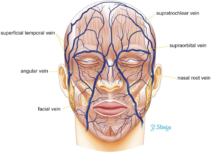
Fig. 7.3 A common pattern of the facial veins. The facial vein begins at the angular vein and descends obliquely near the nasolabial fold.
Cranial to the mandible, the deep facial vein from the pterygoid venous plexus and the inferior palpebral, superior and inferior labial, buccinator, parotid, and masseteric veins join the facial vein. Caudal to the mandible, the submental, tonsillar, external palatine, and submandibular veins join the facial vein.
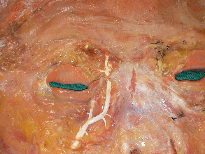
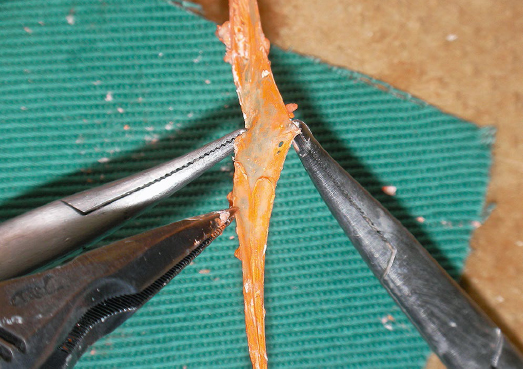
The facial vein has valves, particularly around the level of the mandible (Fig. 7.6).10 The distribution of venous valves indicates that the blood flow is caudal toward the internal jugular vein in the lower part of the facial vein and normally toward the cavernous sinus in the superior ophthalmic vein.6
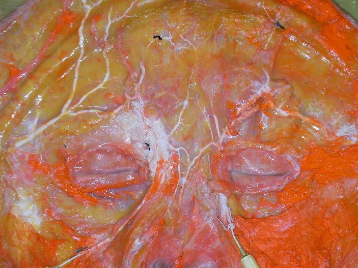
Fig. 7.4 The nasal root vein. The nasal root vein connects the bilateral hemiloop-like veins at the nasal root. Several tributaries from the external nose connect to the vein.
Surgical Annotations
The facial vein connects with the cavernous sinus by two major routes, that is, through the superior ophthalmic vein or through the deep facial vein to the pterygoid plexus and, finally, the cavernous sinus. Thus, infection may spread from the face to the intracranial venous sinuses.
Pterygoid Venous Plexus
The pterygoid venous plexus is an extensive network of small vascular channels that are located between the temporalis and lateral pterygoid muscles (Fig. 7.7). The sphenopalatine, deep temporal, pterygoid, masseteric, buccal, alveolar, greater palatine, and middle meningeal veins and a branch from the inferior ophthalmic vein join the plexus. The plexus connects with the facial vein through the deep facial vein and with the cavernous sinus through the sphenoidal emissary foramen, foramen ovale, and foramen lacerum. Its deep temporal branch often connects with tributaries of the anterior diploic vein and thus with the middle meningeal veins.
Related posts:


Stay updated, free articles. Join our Telegram channel

Full access? Get Clinical Tree


