Chapter 37 Vascularized Tendon Graft for Extensor Tendon Reconstruction
Outline
Composite tissue loss of the hand involving tendon defects represents a great clinical challenge. These injuries require restoration of both skin coverage and tendon function. These injuries are approached by different ways: multistage reconstructions of soft tissue then tendon, or single-stage vascularized composite tissue grafting.1
Multistage reconstructions include skin coverage with distant flaps and tendon grafting in a later stage.2 Nonvascularized tendon grafts in conjunction with pedicled flaps or free tissue transfer are termed partially vascularized tissue transfer.1 Skin,3–8 fascial,9–11 and muscle flaps12,13 can be used for this purpose. A completely vascularized single-stage reconstruction uses a composite flap in which different tissue components (skin, tendon, and nerve) are included.
In 1979, Taylor and Towsend14 described for the first time a composite free flap, with attached vascularized segments of the extensor hallucis brevis to the great toe and the extensor digitorum longus to the second toe. Subsequently, several authors15–19 have described good results for the treatment of compound injuries with the dorsalis pedis composite flap; this flap provides four vascularized tendons (extensor digitorum communis [EDC]) of adequate length.
Reid and Moss20 modified the radial artery forearm flap to include flexor tendons from forearm. The tendons can be included are the palmaris longus (PL) and a strip of the brachioradialis tendons along with fascia and skin, and a slip of flexor carpi radialis (FCR) tendon.21,22 The ulnar island flap of the forearm allows for the inclusion of the PL and a strip of the flexor carpi ulnaris (FCU) tendons.23,24
Operative Techniques
Ulnar Artery–Based Island Flap With Vascularized Tendons (Used by J.C.G.)
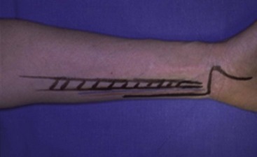
Preoperative evaluation includes the Allen test and Doppler testing to ascertain that the radial artery provides adequate blood supply to the hand. Angiography of the arm is also advisable. A bayonet-shaped incision is first traced and then made on the medial side of the forearm, the axis of the incision overlying the lateral border of the flexor carpi ulnaris (Figure 37-1). The ulnar pedicle is dissected and all its branches are carefully separated and divided.

Figure 37-1 Skin incision for harvesting the ulnar artery-based skin flap incorporating half of the FCU tendon.
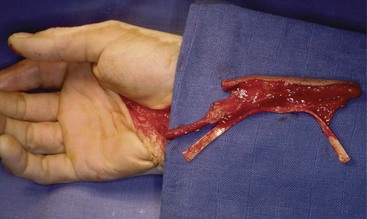
The FCU tendon is longitudinally split into two parts. One is maintained in place with the muscle and the other one (i.e., the medial part) is transected at its distal insertion on the triquetrum. The proximal portion is transected at the musculotendinous junction. All the other branches of the ulnar artery are ligated, except branches to the skin flap on the anterior side and to the periosteum in the event of a bone transfer. The ulnar pedicle is dissected distally and the combined tendon–skin transfer is performed (Figure 37-2).
Postoperative Care
We perform split-thickness skin grafting on the defect immediately following flap harvesting. When the dorsalis pedis flap is used, the ankle, foot, and toes are splinted to prevent movement under the graft. When the radial forearm flap is used, the wrist and the fingers are immobilized for 10 days with the wrist in 20° to 30° of extension with the metacarpophalangeal (MCP) joints in 50° of flexion and the interphalangeal (IP) joints in extension.4
Initially, the hand is splinted with the wrist at 30° of extension and MCP and IP joints in 0°. In our case series, these injuries have been managed with variable periods of immobilization after repair; nevertheless, rehabilitation of the extensor tendon has recently been settled in favor of early mobilization.25


