Molecular Biology of Tendon Healing

Chapter 44 Molecular Biology of Tendon Healing


This chapter does not appear in the print edition.


The weak healing capacity of the injured tendons, intrasynovial tendon in particular, is a critical issue underlying difficulties in achieving optimal outcomes after tendon surgery. After surgical repair, tendons ought to be able to move to prevent adhesions; the weak healing predisposes tendons to rupture during the early tendon movement. In the early half of the 20th century, the mechanisms of healing of primary end-to-end repair of the intrasynovial tendon were not of great concern, because secondary tendon grafting was advocated to treat tendon injuries in the digits. In the 1950s and 1960s, the healing potential of primarily repaired tendons in the sheath area became an essential question facing hand surgeons. In the recent decades, the healing of flexor tendons has been the subject of investigations of a great number of investigators.


In the 1970s and 1980s, a number of elegant experimental investigations demonstrated that cells in the intrasynovial tendon segment can proliferate and participate in the healing process, establishing the concept of “intrinsic tendon healing.”17 Since 1990s, investigations into tendon healing biology have been directed toward elucidation of molecular events underlying the healing process and development of molecular therapeutic methods to enhance the healing or to decrease adhesion formations. These investigations came from several major laboratories around the world, under investigators such as Abrahamsson,810 Chang,1114 Gelberman, Boyer, and Thomopoulos,1520 McGrouther,2124 Mass,25,26 Tang,2734 and Wolfe.35


Currently, laboratory research is focused on uncovering gene expression patterns of the healing tendon and exploring cellular and genetic therapeutic approaches to strengthen weak intrinsic healing capacity. Work is also aimed at improving the healing strength of the surgically repaired tendon.


Two Mechanisms of Intrasynovial Tendon Healing


It is now generally believed that intrasynovial flexor tendons heal through two mechanisms—intrinsic and extrinsic. Both intrinsic and extrinsic tendon healing mechanisms come into play in a clinical setting. Intrinsic healing takes place through proliferation of tenocytes and production of extracellular matrix by intrinsic cells. Extrinsic healing is the healing process brought about by growth of tissues or cell seeding from outside the lacerated tendon. Clinically, the participation of intrinsic and extrinsic healing after tendon repair depends on the condition of the tendon and surrounding tissues. Extrinsic cells participate minimally in the healing of clean-cut wounds but can dominate healing when the intrinsic healing is jeopardized by severe trauma to the tendon.


For a typically lacerated tendon, the healing process generally consists of three stages: inflammation, collagen production (or proliferation and repair), and collagen remodeling.


The first stage is manifested with infiltration of inflammatory cells, such as neutrophils. Monocytes and macrophages are recruited to the injury site within the first 24 hours, and phagocytosis of necrotic materials occurs. After the release of vasoactive and chemotactic factors, angiogenesis and proliferation of tenocytes are initiated. Tenocytes then move into the site and start to synthesize collagen III. The inflammation stage usually lasts for a few days, followed by the collagen production stage.


In the collagen production stage, which lasts for 5 to 6 weeks, the tenocytes produce large amounts of collagen and proteoglycans at the repair site. In the early part of this stage, tenocytes proliferate dramatically, but in later weeks, inflammation subsides and cell proliferation is less obvious. Collagen I, which increases healing strength, is produced abundantly in the later period.


At about 6 weeks after injury, the remodeling stage begins. The first part of this stage is consolidation, which lasts from about 6 to 10 weeks after the injury. During this time, synthesis of collagens and glycosaminoglycans (GAGs) decreases, as does cellularity. The tissue becomes more fibrous as a result of increased production of collagen I, and the fibrils become aligned in the direction of mechanical stress. The final maturation stage occurs after 10 weeks, and there is an increase in cross-linking of the collagen fibrils, which makes the tissue stiffer.


Molecular Biology of Tendon Healing


Roles of Individual Growth Factors


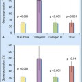
A variety of molecules are involved in tendon healing.36 The roles of six growth factors have been studied associated with tendon healing in vivo or tenocyte proliferation in vitro:


1. Transforming growth factor (TGF). This molecule is secreted by all of cell types participating in the healing process and plays a role in wound healing and scar formation. TGF-β has three isoforms: TGF-β1, TGF-β2, and TGF-β3; they play different roles in scar formation. While TGF-β1 and -β2 are known to induce fibrotic changes and scar formation, TGF-β3 may inhibit scar formation.37 Three isoforms are 60% to 80% homologous and are dimers of 12.5-kDa polypeptides cleaved from larger precursors after being secreted from the cells into the ECM. TGF-β is a master mediator in the pathogenesis of fibrosis and scar formation. Regarding specific roles of TGF-β in flexor tendon healing, Chang and colleagues found increased levels of TGF-β1 mRNA in the sheath and the tendon itself after tendon trauma. TGF-β receptors 1, 2, and 3 were upregulated as well. In in vitro studies, TGF-β1 induced collagen I production by tenocytes.11 Chang and colleagues applied TGF-β1 neutralizing antibodies to injured digital flexor tendons, and this therapy increased the range of digital motion of the toes operated upon in a rabbit model.13

2. Insulin-like growth factor (IGF). It is a single-chain polypeptide hormone. The primary biological action of IGF-1 is mitogenic activity for a broad range of cells. Extracts from the epitenon and internal compartment of avian flexor tendons were found to contain IGF-1; these extracts could stimulate DNA synthesis by tenocytes.8 In in vitro culture of tenocytes, IGF-1 was found to increase cell proliferation as well as stimulate synthesis of DNA, collagen, and proteoglycan.9 Both IGF-1 and IGF-2 were shown to promote proliferation of tenocytes in vitro tenocyte culture.810


Direct injection of VEGF increased the healing strength of Achilles tendon in a rat model. However, the effectiveness of direct use of VEGF in synovial tendons has not been tested. We found that AAV2-VEGF treatment significantly increased the healing strength of the chicken flexor tendons at postoperative weeks 3, 4, and 6.


Both direct delivery of bFGF and transfer of bFGF cDNA via appropriate vectors to the healing tendons have been tested to enhance tendon healing.31,4043 Direct delivery of bFGF was achieved with direct injection of the bFGF,40 use of bFGF-coated suture,41 or bFGF delivery through controlled release system.42,43 Direct injection of bFGF did not improve the healing strength of the tendon, nor did bFGF delivered through controlled release system.40,43 After delivery of bFGF to the tendon, biological reactions and increases in cellular proliferation in the healing tendons were observed, but mechanically, the healing strength was not increased.43 Tendon repair with bFGF-coated surgical suture increased the tendon strength at week 3 after surgery, but not at week 6, in a rabbit model.41 AAV2-bFGF–treated tendons had a significantly greater breaking strength than the nontreatment control at weeks 2, 3, and 4 in a chicken model.31

6. Bone morphogenetic proteins (BMPs). BMPs are a group of growth factors originally discovered by their ability to induce the formation of bone and cartilage. BMPs are considered to constitute a group of pivotal morphogenetic signals, orchestrating tissue architecture throughout the body. There are BMPs 1 through 15 in this superfamily. BMP14 is called growth differentiation factor-5 (GDF-5) as well; this factor was particularly studied regarding its role in tendon healing and repair. Hogan and colleagues44 found that growth differentiation factor-5 (GDF-5, or BMP14) regulates ECM gene expression in murine tendon fibroblasts. They isolated mice Achilles tendon fibroblasts and treated them with rGDF-5 (0 to 100 ng/mL) for 0 to 12 days in cell culture. The temporal effect of rGDF-5 on ECM gene expression was analyzed for type 1 collagen and aggrecan expression. They found that expression of extracellular matrix (ECM) genes procollagen IX, aggrecan, matrix metalloproteinase 9, and fibromodulin were upregulated. Proinflammatory reaction genes were downregulated. rGDF-5 led to an increase in total DNA, glycosaminoglycan (GAG), and hydroxyproline (OHP). rGDF-5 treatment showed improved collagen organization over controls.

No studies have directly investigated expression profile of BMPs during intrasynovial tendon healing process. However, studies were performed to determine relation of BMPs with the healing in rat Achilles tendon. Eliasson and colleagues45

Only gold members can continue reading. Log In or Register to continue

Stay updated, free articles. Join our Telegram channel

Mar 5, 2016 | Posted by in Hand surgery | Comments Off on Molecular Biology of Tendon Healing

Full access? Get Clinical Tree

Get Clinical Tree app for offline access