Chapter 28 Staged Tendon Grafts and Soft Tissue Coverage
Outline
The primary, and most significant, problem of flexor tendon surgery is the formation of adhesions between injured tendons and their surrounds, particularly the tendon sheath within the digits and distal palm. Overcoming adhesion of repairs to the sheath has been the driving force for most research in this field over the past 100 years. Mayer and Ransohoff (1936), writing in the era when delayed flexor tendon grafting was the routine,2 described how “these adhesions extend from the point of division of the tendons down to the distal end of the digital theca (tendon sheath)… . It is obvious that the normal gliding mechanism of the tendon has been completely destroyed and that, consequently, the conditions for re-establishing free gliding of a transplanted tendon are so unfavorable that only in exceptional instances can an implanted tendon perform its normal function.” Mayer was the first surgeon to try to reestablish the milieu required for free gliding of the tendon within the sheath by insertion of an inert rod, then, at a later stage, replace this with a tendon graft. He used celloidin tubes but had to abandon his experiment to create a “pseudo-synovial” sheath as these were too rigid. Twenty-five years later, Bassett and Carroll (1963) repeated the work using silicone rods.3 James Hunter carried this work forward through the 1960s and 1970s to establish the technique clinically.1,4–10 The successors to the rods of woven Dacron covered by silicon developed by him for use in staged flexor tendon grafting are known to us all today simply as “Hunter” rods. Curiously, Hunter’s first use of a silicone rod was to replace an extensor tendon. The rod was sutured to the extensor tendon with fine wire. It became clear that the rod was being stretched and acting as an elastic band to pull the flexed finger back into extension when the flexor tendon relaxed. Had the wire suture not protruded through the skin, there would have been no second stage. On exploration of the protruding wire, it was clear to Hunter that a shiny mesothelium-lined membrane had formed around the rod. The rod was replaced by a tendon graft and the finger went through rehabilitation to achieve acceptable function. Hunter’s initial intention was to design an artificial tendon for permanent use and he pursued both the concept of the pseudo-sheath and the idea of a permanent artificial tendon replacement in his research. While the former has become part of the armamentarium of most hand surgeons, a permanent artificial tendon remains “experimental,” largely because of the problems of achieving a permanent bond between the rod and the biological tissues to which it must be attached proximally and distally.
The Pseudo-Sheath
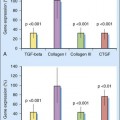
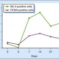
After implantation, the silicone rod holds the adjacent tissues apart. Microscopic studies show that the pseudo-sheath is not simply a tube of scar.11 The tissues adjacent to the rod organize over a few days into a mesothelium-like layer. In the chicken experiments described by Salisbury and his colleagues, this intima consisted predominantly of one layer of flattened fibroblasts with a surface made irregular by pleats and pores, whereas the normal tendon sheath has an intima of several layers of thicker, cuboidal cells. The intima also contained macrophages, intermediate cells, mast cells, and Schwann cells. By 4 to 6 weeks, the deeper tissues have formed a second layer of loose, well-vascularized connective tissue and the pseudo-sheath has a basic appearance similar to normal synovial sheath. The collagen in the outer layer was of variable thickness and contained collagen, reticulum, and elastic and unmyelinated nerve fibers. The collagen was orientated along the axis of the implant much as it is orientated along the lines of stress in a tendon sheath.
Choice between Single-Stage and Two-Stage Grafting
Although much of the flexor tendon surgery performed in western Europe is primary surgery, we do have to undertake a surprising amount of secondary flexor surgery, some of which is because of delayed presentation and some of which is because the flexor tendon injury quite frequently does not come as an isolated and simple one but rather in association with injuries to other tissues. Nevertheless, secondary surgery for us is mostly that of the complications of primary repair, namely ruptured and adherent primary repairs, as the results of the initial surgery are not universally good. In the best units, the failures constitute about 10% of all primary repairs, not a small number and something of an indictment of our present techniques of primary flexor tendon surgery. The cases that come to secondary surgery are mostly either the result of more severe injuries or have occurred in patients who make excessive amounts of scar tissue or have not cooperated with therapy because of low pain thresholds, social circumstances, or lack of judgment.12 Therefore, the cases needing secondary surgery can be considered under the headings of either “bad injuries” or “bad patients.” Elsewhere in the world, many patients will only get to an appropriate surgeon at a time when secondary repair of the tendon using grafts is the only option because of proximal tendon retraction. This, by definition, becomes “secondary flexor surgery,” although the problem is simply an extended finger with good passive but no active flexion but now no longer amenable to direct repair. However, among the patients presenting after delay, and also defined as undergoing “secondary flexor surgery,” are a group with much more complicated problems, sometimes as a result of injuries to the other structures of the digits and, sometimes, as a result of the unaided healing process within a digit in the presence of an inactive flexor system. The problems in these cases are not simply those of being unable to get the tendon ends together. So, in terms of the pathologies in the digits themselves, the problems we all face in the rather heterogeneous mix of cases that we call “secondary flexor tendon surgery” are not so different—namely, flexor tendons that are not intact and flexor tendons that are stuck in scar tissue, variably associated with divided pulleys, skin deficit on the flexor aspect of palm and digits, stiff fingers, and injuries to other, adjacent structures in the fingers, hand, or forearm.
Previously unemphasized reasons for staging grafting have become evident in my practice over the past 20 years. Some patients present with ruptures of primary repairs and undergo immediate exploration with a view to re-repair.13 If it is found at surgery that re-repair is not possible, we routinely put a tendon rod into the finger, with the rod being replaced by a graft when the finger has settled. At this point in time, the finger is often unsuited to a single-stage tendon graft procedure because of its swollen condition, this being the third traumatic episode for this finger (exploration of the rupture following after the causative incident and the primary repair) in a short period of time. Others who rupture a primary repair and cannot undergo immediate re-repair for reasons such as skin breakdown and infection and those who re-rupture the re-repair will present at a later stage for tendon grafting but also may be unsuitable for single-stage grafting, for a variety of reasons, including those grouped together above under “bad injury” and “bad patient.”
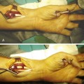
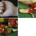
A small group of patients presenting with severe and/or contaminated injuries, sometimes with missing segments of the flexor tendons, are deemed unsuitable for primary repair. This subject is discussed in Chapter 9 by Professor Tang. While we endeavor to carry out primary repairs now in most of these patients while carrying out the other procedures necessary to their overall hand reconstruction, then mobilize them as early as possible, there are some cases where primary repair is impossible. Whenever possible in such cases, we insert tendon rods into the flexor sheaths to maintain these until the flexor tendons can be reconstituted at a later date.

Stay updated, free articles. Join our Telegram channel

Full access? Get Clinical Tree


