This article is a clinically practical review structured around the specific applications of laser technologies used in acute management of soft tissue injuries in surgical incisions and trauma. Surgical and traumatic incisions and injuries provide the clinician with the unique opportunity to follow the progression and maturation of the wound healing response from a very early stage. There has been a recent interest in early cosmetic optimization of surgical and traumatic wounds on the face using optical technologies. Early clinical results for acute laser intervention starting immediately after suture removal or the first several weeks after repair have been very promising.
This article discusses use of laser technologies in acute management of soft tissue injuries in surgical incisions and trauma. To minimize scar formation, current standard of care in acute management of surgical incisions includes irrigation and cleansing, multilayered, tension-free closure with precise approximation and eversion of wound edges, judicious use of suture material, use of postoperative moisture barrier or dressing, and early removal of surgical sutures. Traumatic soft tissue injury involving the skin can be more challenging to manage acutely due to frequent presence of crushed, macerated, or otherwise devitalized tissues. Additional steps are often warranted in that setting including removal of foreign bodies, copious irrigation of the wound, removal of clearly devitalized tissues, and use of antibiotics to cover polymicrobial flora. Despite these measures, poor cosmetic outcome is frequent after surgical procedures and traumatic skin injuries.
Numerous adjunctive measures have been proposed to optimize wound healing and obviate the need for operative scar revision, many of which are discussed in this volume. These include use of steroids, post-treatment dressings, avoidance of sunlight, dermabrasion, and laser treatment. In classic dermabrasion, mechanical debridement of the superficial papillary dermis leads to re-epithelialization via the adnexal structures, resulting in improved texture and color of the skin. This is an excellent method for smoothing an irregular surface or correcting pigmentary discrepancy between adjacent skin edges, which alters how light creates shadows across the surface. Dermabrasion is recommended 6 to 8 weeks after injury/surgical procedure. More substantial improvements are reported during this period, as the immature scar is still undergoing remodeling, rather than during the mature phase. Proposed mechanism of action for this modality has been described by Harmon and colleagues as reorganization of connective tissue ultrastructure and epithelial cell–cell interactions with an increase in collagen bundle density and size with a tendency toward unidirectional orientation of fibers parallel to the epidermal surface. Although excellent outcomes have been described with this technique, it does require a fair level of operator experience and learning curve for manual control of the depth of dermabrasion and feathering of the edges. Additionally, this technique can be complicated with excessive bleeding and tearing of tissues at the treatment margin. Laser scar revision is a competing technology that has gained increasing popularity due to the potential for excellent hemostasis, ease of use, and precise control over depth of penetration and extent of treatment.
Since the introduction of laser skin resurfacing for aesthetic surgery in the mid-1990s, the technology has worked its way into broad use in scar revision. Laser and optical methods for management of acute injury have very vital roles in postsurgical and traumatic wound therapeutic outcome. With advances in this technology a clinician may arrive at a crossroads in the decision to treat a scar with surgical revision versus dermabrasion or various laser technologies. The authors present and review their experience herein to help with this decision-making process in the acute setting. This article discusses indications and strengths of available optical modalities with a focus on acute and subacute skin injuries. The discussion is practically oriented and structured around the specific applications of each technology.
Acute cosmetic management of surgical wounds
Scars can be disfiguring, aesthetically unacceptable, and cause pruritis, tenderness, pain, sleep disturbance, anxiety, and depression in postsurgical patients. Regardless of the specific strategy for treatment, current optical technologies offer the reconstructive surgeon valuable tools to lessen the psychological burden of undergoing surgery by optimizing the cosmetic outcome in a noninvasive or minimally invasive form. Availability of these tools can lead to higher levels of patient satisfaction after surgery.
For purposes of this discussion, a surgical incision is defined as an incision that is closed per standard of care as discussed previously with optimal postsurgical care and without perioperative wound complications. Ideal post-surgical scar is flat, flexible and indiscernible from surrounding skin in terms of color and texture. Despite optimal wound closure and postoperative care, aberrant fibroblast response can lead to hypertrophic or keloid scars, and aberrant angiogenesis may lead to telangiectasias or a hyperemic scar. Imperfect surgical closure or poor postoperative management can lead to poor outcomes with step-offs, depressions, suture marks, dyspigmentation, or broad hypertrophic scars due to wound tension or distal flap vascular compromise and tissue necrosis. Abnormal collagen deposition has been demonstrated histologically in hypertrophic scars with elevated levels of collagen 3 Traditionally improved via mechanical dermabrasion, the current arsenal for optimization of surgical wounds includes various optical technologies such as conventional ablative laser resurfacing and nonablative laser treatment, as well as fractionated and pulsed laser technologies. Acute optimization of wound healing can start immediately after the completion of surgery as in laser-assisted scar healing (LASH) or after removal of sutures within 1week postoperatively, or it may focus on treatment of maturing scar several weeks to months after surgery.
Acute cosmetic management of traumatic wounds
Some injuries such as uncomplicated linear lacerations are indistinguishable from surgical incisions. The challenge with traumatic skin injuries lies in irregular borders, high tension, macerated tissue, and tissue loss. More often than not, traumatic lacerations involve nonlinear or stellate disruption of the epidermis that is not at right angle to the skin surface. Traumatic abrasions can harbor foreign bodies, which if not adequately debrided, can lead to traumatic tattooing. Presence of tissue edema, hematoma, and loss of tissue can force a high-tension closure, which can lead to broad hypertrophic scarring. Risk of infection is compounded by inadequate cleansing and irrigation of tissues and lack of proper antibiotic coverage after repair. Devitalized, necrosed skin edges and infected wounds can lead to severe atrophic or hypertrophic scars and extremely poor cosmetic outcomes. Use of copious irrigation and antibiotic coverage for gram-positive skin flora with first- or second-generation cephalosporins or clindamycin should be considered in these patients. In the case of an animal or human bite or other gross contaminations of the wound, appropriate adjustments to the antibiotic coverage must be made.
Due to the specific patient population as well as the treatment setting, poor follow-up is often an issue with these patients, leading to improper postoperative care such as suture retention or delay in diagnosis of wound infection. Use of rapidly absorbing suture material where available and appropriate is therefore advised in traumatic skin closure, particularly in those whose attention to follow-up is uncertain. Delayed presentation to the surgeon can also be an issue in this population, as the patient is often acutely managed by an emergency room physician, family doctor, physician assistant, or a general surgeon as opposed to a specialist with reconstructive surgical expertise. Intervention should occur as early as possible following presentation to prevent progression in the direction of an undesirable mature scar that may require surgical revision.
Optical management of acute surgical and traumatic wounds
This discussion encompasses conventional ablative resurfacing lasers, pulsed dye laser (PDL), and fractionated lasers for acute cosmetic optimization of surgical and traumatic wounds.
Ablative Laser Resurfacing
Traditional laser resurfacing is a technique that is commonly accomplished via ablative devices such as conventional carbon dioxide (CO 2 ) or Erbium:YAG lasers. Mechanism of action is similar to using a mechanical dermabrader with the potential to modulate wound healing through thermal effects of laser and trigger the same regenerative mechanisms as when these devices are used for classic facial resurfacing. Tissue removal by laser is a function of treatment parameters, tissue optical properties, and tissue thermal properties. Histologically, laser-treated skin shows a subepidermal dermal repair zone consisting of compact new collagen fibers overlying collagen with evidence of solar elastosis. Because it is possible to achieve discrete, measurable incremental amounts of tissue removal with each pulse, only a modest level of skill is required for achieving optimal results.
CO 2
The CO 2 laser is the work horse of cosmetic dermatology and the model to which all optical therapies are compared. Despite nearly 20 years of use, CO 2 laser skin resurfacing remains very valuable due to the capacity to remove bulk amounts of tissue in a bloodless fashion, and correct iatrogenic contour irregularities. Differences of outcomes between CO 2 and dermabrasion remain incompletely understood. However, due to decreasing technology associated costs, CO 2 laser resurfacing has slowly gained popularity. CO 2 laser is emitted at wavelengths ranging from 9400 to 10,600 nm, and it is preferentially absorbed by water (its principal chromophore), leading to superficial ablation of tissue by vaporization. Although the majority of the energy is absorbed by the first 20 to 30 μm of the skin, the zone of thermal damage can be as much as 1 mm deep. This is in part responsible for the persistent erythema experienced by patients that can continue for 6 months or longer after CO 2 laser treatment, but it may also contribute to enhanced collagen remodeling. Timing of the treatment is typically the same as mechanical dermabrasion, optimally performed 4 to 8 weeks after the initial injury. The ideal application of this laser is for induction of contour changes and collagen remodeling in elevated scars.
Erbium:YAG Laser
Introduced to dermatology in the mid-1990s, the Erbium:YAG laser also removes tissue, but the penetration depth of the wavelength (2936 nm) is shallow, as is the corresponding depth of thermal injury. Light from this laser is absorbed 12 to 18 times more efficiently by water compared with the CO 2 laser. However, the more superficial depth of penetration and surrounding tissue injury lead to decreased induction of collagen remodeling and contraction. Due to poor coagulative properties, hemostasis can also be a problem with this modality, particularly if extensive tissue needs to be removed. Application of this laser is for generating subtle contour changes in depressed and atrophic scars. Additionally, the Erbium:YAG laser may be used in cases where thermal injury is undesirable (such as a known keloid former).
PDL
PDL relies upon concept of selective photothermolysis. The 585 to 595 nm wavelengths are preferentially absorbed by hemoglobin, although epidermal melanin absorption can be of concern in patients with darker skin phototypes. This selectivity makes this technology ideal for in the treatment of vascular lesions skin lesions such as telangiectasia, port wine stains, and hemangiomas. During scar treatment, PDL destroys the blood supply to the wound edge at the level of dermal microvasculature, inhibiting the formation of scars. The angiolytic mechanism of action has been disputed by some authors. Alternatively, changes in cell cycle distribution of fibroblasts in keloid scars has been proposed recently as a mechanism of action of PDL treatment in keloid scars.
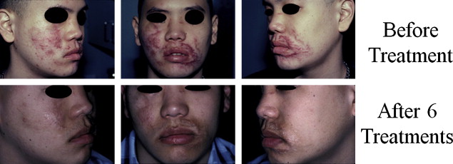
Properties of this laser make it suitable for treatment of red, hyperemic, hypertrophic scars, and keloids ( Figs. 1–3 ). PDL improves color, texture, and pliability of scars by reducing pigmentation, vascularity, and bulk of scar tissue. Because it spares the epidermal and dermal tissues, treatment can be repeated at 6- to 8-week intervals with significantly reduced downtime and erythema compared with conventional CO 2 laser resurfacing. Due to competitive absorption of the emitted energy by melanin, darker toned individuals (Fitzpatrick IV-V) may not be suitable candidates for this treatment due to risk of dyspigmentation. The authors’ parameters for PDL wound optimization are listed in Table 1 .
Related posts:
 Use of Makeup, Hairstyles, Glasses, and Prosthetics as Adjuncts to Scar Camouflage
 Enhancement of Facial Scars With Dermabrasion
 Skin: Histology and Physiology of Wound Healing
Scar Revision Techniques: Z-Plasty, W-Plasty, and Geometric Broken Line Closure
Skin: Histology and Physiology of Wound Healing
Scar Revision Techniques: Z-Plasty, W-Plasty, and Geometric Broken Line Closure
 Enhancement of Facial Scars With Dermabrasion
Laser Treatment for Improvement and Minimization of Facial Scars
Enhancement of Facial Scars With Dermabrasion
Laser Treatment for Improvement and Minimization of Facial Scars
Stay updated, free articles. Join our Telegram channel

Full access? Get Clinical Tree


