Upper eyelid reconstruction with Cutler-Beard flap and free tarsal graft


Table 43.1

Indications for surgery


Reconstruction of large (>80%) full thickness defects of the upper eyelid


Table 43.2

Preoperative evaluation


Size and dimensions of upper eyelid defect
Involvement of lacrimal system
Eyelid laxity
Evaluation of donor sites for grafts (contralateral upper eyelid, ipsilateral lower lid, pre/retroauricular, supraclavicular, inner arm)
History of prior eyelid, facial surgery or trauma
Visual function of both eyes (monocular status)
History of smoking/tobacco use


Introduction


Total defects of the upper eyelid are often the most challenging to reconstruct. Compared to the lower eyelid, the upper eyelid has greater vertical excursion, function and dynamic movement. Ideally, the reconstructed upper eyelid should share a similar purpose with optimum aesthetic appearance.


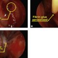
Complete loss of the upper eyelid can occur due to cutaneous malignancy, trauma or developmental anomalies. In reconstruction of larger defects, the bi-lamellar architecture of the eyelid typically requires a combination of flaps and grafts for the most ideal result. Time-honored grafting principles apply in that either the anterior or posterior lamella must provide the vascular supply. Free grafts can be layered on a pedicle-based flap. If the lacrimal system is involved, canalicular reconstruction should be performed primarily but creation of a nasolacrimal conduit should be deferred in cases of tumor.


Smaller defects of the upper eyelid can be closed using a variety of techniques. Direct closure ( Chapter 37 ) and the Tenzel semicircular flap ( Chapter 39 ) work well as non-lid-sharing techniques, but greater sized defects may require pedicle-based grafts from the lower eyelid. Direct closure and the semicircular flap have the advantage of preserving some eyelashes. The Cutler-Beard procedure was originally described in 1955 for reconstruction of large upper eyelid defects. The procedure involves advancing a full thickness (from skin to conjunctiva) myocutaneous flap from the lower eyelid harvested inferior to the inferior tarsal border. The flap is then transferred posteriorly to the preserved lower eyelid margin segment to reconstruct the upper eyelid. Because there is no tarsus present in the advancement flap, a free posterior lamellar graft is typically harvested and placed first in the upper eyelid defect. Choices for posterior lamellar grafts include free tarsal grafts, nasal chondral mucosa and hard palate. A posterior lamellar graft can be omitted but the upper eyelid may be less stable.


As an eyelid sharing procedure, the Cutler-Beard flap needs to gain vascularity prior to the staged release of the pedicle. Ideally this should occur from 4 to 6 weeks after stage one. In monocular patients and in children, eyelid-sharing techniques should be used cautiously due to the interference with vision and possible amblyopia.
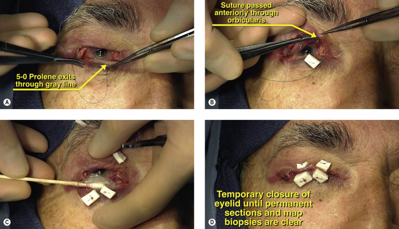


Surgical Technique


Figures 43.1A and 43.1B


Incisional biopsy of eyelid

This 82-year-old man was referred with a non-resolving chalazion of the right upper eyelid. On examination, the lesion appeared as a firm, yellow mass disrupting the eyelid margin and with associated madarosis ( Figure 43.1 ). A full-thickness biopsy of the lesion revealed sebaceous cell carcinoma ( Figure 43.2 ). Sebaceous cell carcinoma occurs more frequently on the upper eyelid and presents classically as chronic unilateral blepharitis or chalazion, as in this case.


Figures 43.2A–F


Wedge excision of eyelid lesion

For sebaceous cell carcinoma and melanoma of the eyelid and periorbita, a surgical margin of at least 5 mm should be obtained ( Figure 43.2A ). Vertically, the entire tarsal plate should be removed also with a margin of at least 5 mm ( Figure 43.2B ). The vertical eyelid incisions are made with sharp iris scissors ( Figures 43.2C and 43.2D ). At the superior tarsal border, the incision is made with Westcott scissors to remove the entire eyelid wedge ( Figure 43.2E ). The tissue is then placed in formalin for permanent section and all instruments used during the excision are removed from the surgical field to prevent tumor dissemination ( Figure 43.2F ).


Figures 43.3A–D


Map biopsy of bulbar and palpebral conjunctiva

Sebaceous cell carcinoma can exhibit Pagetoid or skip lesions with intervening normal areas of tissue. Map biopsies of the conjunctiva survey the entire ocular surface to determine if local metastasis is present. The bulbar conjunctiva is sampled at six areas around the limbus, as shown in Figure 43.3A . The palpebral conjunctiva and tarsus are sampled at four areas, as shown in Figures 43.3B and 43.3C . Each individual specimen can be sent in a separate formalin container wrapped in non-absorbable gauze (Telfa pad) or placed on a sterile sheet of cardboard with the surgical margins marked ( Figure 43.3D ). All margins should be sent for rush paraffin-embedded permanent sections. Frozen sections are not sufficient to rule out residual tumor in either sebaceous cell carcinoma or melanoma.


Figures 43.4A–E


Advancement of conjunctiva

The cut edge of the conjunctiva is identified and this is advanced to the skin edge to preserve the cul-de-sac during reconstruction ( Figures 43.4A and 43.4B ). Interrupted 7-0 Vicryl sutures are used to advance the conjunctiva to prevent retraction deep into the fornix during the postoperative period ( Figures 43.4C and 43.4D ). Examination of the upper eyelid shows a greater than 80% defect ( Figure 43.4E ). Once all margins have been cleared by permanent sections, the upper eyelid can be reconstructed. If the margins are positive, a wider excision is performed with repeat permanent sections.

May 16, 2019 | Posted by in Reconstructive surgery | Comments Off on Upper eyelid reconstruction with Cutler-Beard flap and free tarsal graft

Full access? Get Clinical Tree

Get Clinical Tree app for offline access