Reconstruction of the upper eyelid is complicated because the eyelid must retain mobility, flexibility, function, and a suitable mucosal surface over the delicate cornea. Defects of the upper eyelid may be due to congenital defects or traumatic injury or follow oncologic resection. This article focuses on reconstruction due to loss of tissue. Multiple surgeries may be needed to reach the desired results, addressing loss of tissue and then loss of function. Each defect is unique and the laxity and availability of surrounding tissue vary. Knowing the most common techniques for repair assists surgeons in the multifaceted planning that takes place.
Key points
- •
In eyelid wound closure, tension should be drawn horizontally to avoid ectropion or lid retraction.
- •
Local myocutaneous advancement flaps may be used for smaller defects. For moderate to large defects, the Tenzel semicircular advancement flap technique or the Cutler-Beard 2-staged procedure may be used, respectively.
- •
When the lid margin is involved, eyelash growth may be compromised. Loss of the medial eyelid margin is further complicated by the lacrimal drainage system and medial canthal tendon attachment to the lacrimal crest.
- •
Understanding the factors of upper eyelid reconstruction and knowing different techniques for repair prepare the surgeon to create a functional and cosmetically acceptable eyelid.
Introduction
A patient undergoing reconstructive surgery of the eyelids desires results that best restore the eyelid function while optimizing the aesthetic outcome. Reconstruction of the upper eyelid is more challenging than that of the lower eyelid due to the highly specialized function of the upper eyelid. The eyelid is a thin structure with the ability to open and close to lubricate and protect the ocular surface while allowing an unobscured visual axis to see. The maintenance of the corneal surface is critical for sharp vision and patient comfort. Any disruption of the eyelid tissue can cause loss of mobility of the eyelid by scarring and tethering of the lid margin. Loss of tissue is particularly troublesome because the eyelid must maintain its mobility, flexibility, function, and a good mucosal surface to be in contact with the sensitive cornea. Dynamic blink and range of movement is often at odds with the need to replace tissue to allow for the eyelid to close. Inability to close the eyelids together can cause drying of the eye, which leads to blurred vision, light sensitivity, and increased risk of infection that could eventually lead to loss of the eye. Multiple surgeries may be required to achieve the desired results, to address first the loss of tissue and then the loss of function. In addition, the surgeon must also take into consideration the appearance of the eyelid, which can have an adverse impact on a patient’s quality of life.
Defects of the upper eyelid may be related to various causes, including but not limited to congenital defects and traumatic injury, or follow oncologic resection. The type of reconstruction depends on the anatomic deficits identified, including the vertical, horizontal, and depth dimensions as well as the availability of regional and distant tissue for reconstruction. This article focuses on reconstruction techniques used in the correction of eyelid defects that involve loss of tissue.
Anterior lamellar upper eyelid defects
The anterior lamella of the eyelid includes skin and orbicularis oculi muscle. Small defects less than 1 cm in diameter can heal well by secondary intention if they are superficial and favorably located in the medial canthus, which has bony support, or along the pretarsal skin, in which the tarsus lends support to the wound. Contracture of the wound is typically noted by 2 to 6 weeks, at which time the need for further surgery can be determined for possible sequelae, such as lid retraction or ectropion of the eyelid margin.
When there is redundant skin adjacent to the defect, primary closure is often possible with or without undermining the skin. The ability to close without a formal flap depends on the quality of the skin and the amount of redundancy present. Closure should draw the tension horizontally to avoid ectropion or lid retraction, although aligning scars with the lid crease can help camouflage the scar.
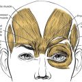
When more tissue recruitment is needed, local myocutaneous advancement flaps are the preferred choice because they provide optimal color and texture match, near-normal innervation, minimal contracture, and a rich blood supply. The most useful flaps in this area include rhombic, transposition, and advancement flaps ( Fig. 1 ).
When the lid margin is involved, the anterior lamella contains eyelashes, which may be lost or may regrow through scar tissue, causing trichiasis. Special consideration should be made if the eyelashes are involved because this marginal tissue is adherent to the underlying tarsus and does not allow for direct closure. The upper eyelid eyelashes are more visible than the lower eyelid lashes and maintaining a continuous lash line can be an important part of the final cosmesis. Converting this defect into a full-thickness defect can be a viable and preferred option when there is less than 33% of the lash margin loss. The lack of lashes laterally is not as noticeable because it is centrally and there seems to be a compensatory eyelash follicle mechanism that maintains the total number of eyelashes.
Skin grafts offer replacement of the eyelid skin with tissue taken from a distant source when local options are exhausted. A full-thickness skin graft is preferable to a split-thickness skin graft due to less wound contracture. Optimal donor sites have thin dermis, minimal hair, and similar color and texture to the eyelid. Contralateral upper eyelid skin is the best match, followed by lower eyelid skin, preauricular or postauricular skin, supraclavicular skin, and inner upper arm skin. The color match from these sites has been found to be good in 85% to 94% of patients, with more hypopigmentation noted more from supraclavicular and inner upper arm donor sites. Complications of periocular full-thickness skin grafts occur in approximately 12% of cases. The most common complications were hypertrophy (42.3%), partial graft failure (27.2%) and wound contraction (15.3%).
A split-thickness graft allows for significantly more coverage of a wound because the donor site heals by secondary intent and the hair-bearing skin may be used as the follicles are left behind. The contracture of a split-thickness graft can cause eyelid retraction and compensatory eyelid tightening or a temporary tarsorrhaphy may be needed to reduce this effect during the healing phase. Massage, steroid ointment or injections, and silicone gel application can also be useful in the postoperative phase to improve hypertrophy or contracture of the skin grafts.
Full-thickness upper eyelid defects
Canthal Defects
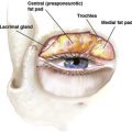
Loss of the medial eyelid margin is complicated by the presence of the lacrimal drainage system and the medial canthal tendon attachment to the anterior and posterior lacrimal crest. Reconstruction of the lacrimal drainage system typically involves silicone stent intubation of the remaining canalicular system to maintain patency. Although the upper eyelid is not as critical in tear drainage as the lower eyelid is, delayed repair can be difficult to achieve with excellent success rate of primary repair.
Identifying a suitable anchoring point for the medial upper eyelid is important for stabilization of the lid margin. Optimal choices include the stump of the medial canthal tendon or the periosteum of the medial orbital wall. Microplates, miniplates, resorbable and nonresorbable screws, and transnasal wiring have been used when insufficient soft tissue remains.
Loss of the lateral canthal tendon may be replaced by creating a lateral tarsal strip or a periosteal flap from the lateral orbital rim. It is best to create an oblique strip of periosteum hinged at the orbital rim that is longer than believed needed. The lateral orbital rim provides the ability to drill a full thickness hole for direct anchoring to the bone with suture or wires if there is not enough soft tissue.
Small Defects
Depending on the laxity of the eyelid tissues, full-thickness defects of the lid margin involving up to one-third of the horizontal length may be closed with direct closure. Assessment includes pulling the edges of the defect together to confirm the wound is not under too much tension. Minimal tension can be released with a lateral canthotomy or cantholysis. If superior tarsus remains, completion of a pentagonal resection or complete excision of the residual tarsus may be performed to allow for the ends to meet appropriately. If there is minimal tarsal loss with good skin laxity, then a local tarsoconjunctival flap may be used to recreate the posterior lamella with skin or myocutaneous advancement flap. Approximately 4 mm of vertical height in the residual tarsus is needed to maintain stability of the lid margin. As discussed previously, the loss of eyelashes can be aesthetically displeasing but preferable if tissue recruitment from the lateral canthal angle is difficult.
The steps of eyelid margin repair include
- 1.
Reforming the lid margin: use a 5-0 or 6-0 Vicryl suture on a spatulated needle to place partial-thickness pass through the tarsus. Properly align the lid margin by positioning the suture obliquely through the tarsus with the anterior pass level with the base of the eyelashes and the posterior pass level with the corner of the lid margin. Maintain partial-thickness passes to avoid corneal abrasion ( Fig. 2 A).