Lower eyelid defects are common, and a systematic approach to reconstruction of the lower eyelid is required. Attention to the bilaminar eyelid anatomy and canthal support structures, with efforts to maintain functionally important structures, such as the lacrimal canalicular system, is vital to appropriate lower eyelid reconstruction. Techniques of advancement and rotation flaps and grafting of skin and mucosa are mainstays of lower eyelid reconstruction. An appropriate armamentarium of techniques allows for optimal surgical results.
Key points
- •
The eyelid has an anterior lamella of skin and muscle and a posterior lamella of mucosa that must be reconstituted in any reconstruction.
- •
Composite flaps or an anterior or posterior lamellar flap and opposing graft may be used in reconstruction of the eyelid margin.
- •
The medial and lateral canthi are vital supportive structures and maintain the shape and position of the eyelid. Their function must be maintained in eyelid reconstruction.
- •
Whenever possible, the function of the lacrimal canalicular system must be maintained or restored in eyelid reconstruction.
- •
Surgeons must possess a range of reconstructive options to provide optimal results in eyelid reconstruction.
Introduction
Knowledge of anatomy and function of the eyelids is essential when faced with the challenges of eyelid reconstruction. Trauma can cause significant tissue loss, which necessitates reconstruction, rather than primary closure, and correcting congenital defects may also require reconstruction and repair. Most commonly, periocular reconstruction follows excision of malignancies. An aesthetically optimal restoration of anatomy and function is always the paramount goal in eyelid reconstruction.
Anatomy
The eyelids protect and lubricate the ocular surface. The tear film and corneal interface is essential for vision. An inadequate tear film causes blurred vision, infection, and scarring, or even loss, of the eye. With each blink, tears coat the cornea, providing lubrication, essential nutrients, and oxygen to the ocular surface.
The eyelid is made up of anterior and posterior lamellae. The posterior lamella of the eyelid is lined with conjunctiva, a nonkeratinized epithelial mucous membrane filled with secretory glands. Anterior to and intimately attached to the conjunctiva at the lid margin are the more rigid tarsal plates composed of dense connective tissue that provides support to the eyelids. The tarsus has its largest vertical height centrally (10–12 mm upper eyelid and 4 mm lower eyelid) and tapers toward the canthal angles attaching to the canthal tendons.
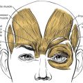
The anterior lamella consists of the skin and muscle of the eyelid. The skin of the eyelid is unique because it is the thinnest in the body. It is devoid of subcutaneous fat, so the epidermis and dermis directly overlie the orbicularis oculi muscle. The orbicularis oculi is anterior to the tarsal plates and deep to skin. The orbicularis oculi provides for eyelid closure. Forceful closure is obtained from the orbital portion of the muscle, whereas involuntary blink originates from the preseptal and pretarsal portions of the orbicularis oculi. Maintaining orbicularis innervation from the facial nerve is essential to ensure eyelid closure and blink.
The medial and lateral borders of the eyelids are formed by the canthal tendons. The canthal tendons add support to the eyelids and maintain the palpebral fissures. The medial canthal tendon has both anterior and posterior arms that attach to their respective lacrimal crests, encircling the lacrimal sac. Laterally, the canthal tendon attaches above the lateral orbital tubercle, within the orbital rim. The lateral canthal tendon generally attaches 2 mm higher than the medial canthal tendon.
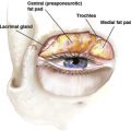
The lacrimal drainage system originates in the eyelids. In the upper and lower eyelids, a punctum is present in the margin, which is the opening to the canaliculi. The first 2 mm of the canaliculi travel vertically from the puncta, before a 90° turn. The canaliculi then run horizontally for 8 mm to 10 mm, beneath skin toward the medial canthal angle. The canaliculi either directly connect to the lacrimal sac or join as a common canaliculus prior to entering the lacrimal sac. The lacrimal sac drains through the nasolacrimal duct beneath the inferior turbinate in the nose.
General principles of eyelid reconstruction
The end goal of any eyelid reconstruction is an aesthetically optimal restoration of anatomy and anatomic function with a minimum of surgical morbidity. Attention to reconstitution of the bilamellar eyelid structure is essential. The surgeon must create an anterior lamella of adequate and aesthetically appropriate skin, preserving dynamic orbicularis muscle function whenever possible, and create a posterior lamella replacing tarsus at the eyelid margin and providing for a smooth mucosal surface that protects and preserves the cornea.
A preoperative consultation should take place whenever possible so the surgeon can plan an appropriate operative environment and anesthesia and can counsel the patient regarding the probable and extreme possible sizes of the surgical defect and the range of surgical techniques that may have to be used in the repair. Possible sites from which flaps and grafts are to be obtained and the potential for morbidity, the need for temporary eyelid closure, secondary procedures, and risks, such as canalicular obstruction and postoperative tearing, are best explained before tumor resection.
Lower eyelid defects are categorized by amount of full-thickness eyelid defect (as percent of eyelid margin), degree of nonmarginal eyelid involvement, total size of defect, canthal involvement, and lacrimal involvement. Knowledge of an array of reconstructive options allows for planning of the surgical repair.
Many lower eyelid reconstructions are accomplished with a bilamellar advancement of anterior and posterior lamella as a composite flap, most frequently from the lateral canthus. This technique is described in detail later. Defects that require a flap to reconstitute 1 lamella of the eyelid generally use a graft to reconstruct the opposing lamella. It is usually impossible to place a graft on a graft, because there is no blood supply to either graft, and both layers undergo necrosis. The surgeon should consider past or future treatments that may compromise repair, including prior surgery in the area and radiation therapy.
Surgical tension in lower eyelid repair should be directed with tension on flap pedicles directed horizontally, most often superolaterally. Any vertical tension on flaps or vertical recruitment of tissue generally results in retraction of the lower eyelid, which is very hard to correct later.
Appropriate medial and lateral canthal fixation is essential to maintain the position, contour, and horizontal dimension of the eyelid. Direct suturing to periosteum, periosteal flaps, and screw or drill hole fixation are often required to stabilize the position of the reconstructed eyelid and maintain an appropriate eyelid fissure and position. In the medial canthus, the presence of the lacrimal drainage system may necessitate its reconstruction or its sacrifice and later Jones tube (conjunctivodacryocystorhinostomy [CDCR or Jones tube]) surgery.
Nonmarginal eyelid defects can still compromise eyelid function, the position of the eyelid margin, and require careful consideration and reconstruction along the general principles of facial reconstruction. Complex defects may extend beyond the surgical skills of a single surgeon and necessitate a multidisciplinary approach to repair.
Eyelid defects and reconstruction
When encountered with full-thickness eyelid defects, replacement of the anterior and posterior lamellae is required for adequate function. Marginal eyelid defects can be classified by the percent of the eyelid margin involved as small (<20%–30%), medium (30%–50%), or large (>30–50%). A range is applied in this classification to make the point that eyelid laxity, condition of the skin, and prior surgery may convert a small (ie, easily repaired) defect in an older patient with skin and eyelid margin laxity to a medium category defect in a young patient or patient with very tight eyelids, tight skin, or prior surgery.
Table 1 shows a simplified scheme for reconstruction of marginal lower eyelid defects. Familiarity with the limits of each surgical technique allow a surgeon to best choose a reconstructive approach. Special situations that may modify the reconstructive technique include medial and lateral canthal involvement and shallow versus deep defects.
| Full-thickness Defect Size | Primary Techniques | Adjunct Techniques |
|---|---|---|
| Small (20%–30%) | Direct closure | — |
| Canthotomy with closure | — | |
| Lateral advancement flap | — | |
| Medium (30%–40%) | Lateral advancement flap | Canthal fixation Canalicular reconstruction Periosteal flap |
| Large (>40–50%) | Lateral advancement flap | — |
| Hughes tarsoconjunctival flap | FTSG | |
| Mustarde cheek flap | Autogenous tarsal graft or hard palate mucosal graft Medial canthotomy/cantholysis |
In some instances, such as a marginal laceration or a small vertical defect, the eyelid can be repaired by direct closure. Care should be taken to not simply pull together larger defects in this fashion, as the lateral canthus is pulled medially with rounding and distortion of the eyelid fissure. The goal of this repair is reapproximation of the eyelid, with a continuous lash margin, which provides optimal functional and cosmetic outcome. The technique of marginal eyelid repair is described later.
Defects involving 30% to 50% of the eyelid require advancement and rearrangement of neighboring tissues, sometimes with grafting, to adequately reconstruct the eyelid. The lateral advancement flap is the go-to approach for moderate-sized lower eyelid defects, and it is a basic technique that all surgeons performing eyelid reconstruction must be familiar with.
Lateral advancement flaps
- 1.
The lateral canthal region and eyelid defect are infiltrated with local anesthetic with epinephrine. This repair can be performed under local anesthetic alone or with monitored sedation or general anesthesia.
- 2.
Prepare and drape the surgical sites; place corneal protectors.
- 3.
Mark a lateral canthotomy from the canthal angle 10 mm to 15 mm superior, then lateral from the canthal angle ( Fig. 1 ). Make the skin incision with a #15 blade and open through the orbicularis muscle with Westcott scissors.
Fig. 1
A 50% full-thickness lower eyelid defect (once tarsal edges are squared) showing superior, then lateral, incision.
- 4.
Undermine and release deep to orbicularis, taking care to not disturb the orbicularis muscle and skin of the flap.
- 5.
Perform a cantholysis by strumming the tendon with Westcott scissors for identification, then incising the tendon. Be sure the tendon is fully released to allow advancement of the eyelid ( Fig. 2 ).
Fig. 2
Release of the lower limb of the canthal tendon leaving the overlying skin and muscle undisturbed.Related posts:

Stay updated, free articles. Join our Telegram channel

Full access? Get Clinical Tree