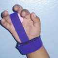
Fig. 10.1
(a) Anteroposterior and (b) lateral radiographs of a 2-year-old boy with bilateral ulnar longitudinal deficiency type II/A. (c) Clinical photograph showing excessive wrist ulnar deviation. (d) The wrist position rests in neutral
Wrist
Angulation of the wrist can occur, but is typically not as severe as seen in radial longitudinal deficiency. (Fig. 10.1c). El Hassan et al. [17] reported that the wrist was positioned in neutral in 71 % of patients, with the remaining wrists resting in 5–40° of ulnar deviation. Those patients with the wrist in neutral position had essentially normal wrist range of motion. However, when the wrist was in ulnar deviation, limitations were documented with regard to radial deviation, wrist flexion, and extension [17]. Controversy over the role of the ulnar anlage and the amount of wrist deviation exists [15–17]. Carpal bones can be absent in correlation with missing digital rays, and synostoses can occur in 30–40 % of cases [15].
Hand
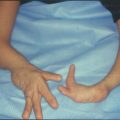
Approximately 90 % of hands with ULD have missing digits and 30 % have syndactyly [1]. Multiple digital anomalies can be seen with the hand ranging from a full complement of digits to just one digit. Ectrodactyly has been well documented in patients with ULD [1, 13, 15, 17]. Many of the existing digits are usually not normal, with variations of hypoplasia, missing phalanges or metacarpals, and variations of syndactyly and synostoses between phalanges and metacarpals [15].
Seventy percent of patients with ULD have abnormalities related to the thumb [1]. El Hassan et al. [17] reported that 11 of 17 limbs with ULD had digital anomalies, with four of those limbs having absent thumbs. Swanson et al. [11] and Broudy and Smith [21] reported that 68 and 100 % of their patients with ULD had radial-sided hand abnormalities, respectively. Cole and Manske [13] reported that 73 % of the 55 hands evaluated had an abnormal thumb or first web space. Their classification system describes the spectrum of thumb and first web space involvement from normal all the way to aplastic [13]. Evaluating the thumb and first web space deficiencies is important because surgical intervention to alter the radial-sided abnormalities in the hand may provide more substantial functional gains than operations elsewhere along the arm [1, 13, 15–17].
Treatment
Treatment of patients with ULD depends on the function of the limb. Non-operative intervention typically consists of early stretching and splinting starting at a young age. Depending on the function of the hand, surgical intervention may be warranted. Thus, the majority of surgical interventions in patients with ULD are performed on the hand, including syndactyly releases, deepening of the first web space, and first metacarpal rotational osteotomy [1, 13, 15, 16]. In special circumstances, other procedures including excision of the ulnar anlage, humeral rotational osteotomy, and creation of a one-bone forearm may be indicated.
Hand
Hand function can be improved with syndactyly releases, reconstruction of the thumb (opponensplasty, pollicization), and deepening of the first web space [1, 16, 23]. Ezaki and Carter [16] recommend delaying hand surgery until the second year of life. The reconstruction procedures of the hand are very important in improving the function of these children, and they recommend waiting for the hands to get larger to allow for a more precise surgery and thus better result [16].
First metacarpal rotational osteotomy is indicated in hands where the digits all lie in the same plane. The goal of rotation is to allow for prehension with the pulp of the digits. Rotation of other metacarpals and even phalanges to achieve this goal should be performed. Ezaki and Carter [16] report that there is a tendency for a slow loss of rotation after surgical intervention, and they recommend concomitant realignment of muscle power with tendon transfers to help prevent derotation.
Wrist
Controversy over excision of the ulnar anlage has been debated within the literature [11, 15–17, 21, 22]. However, there is some agreement as to which patient may benefit from early anlage excision. Indications for ulnar anlage excision [1, 15–17, 22] include the following:
1.
Greater than 30° of fixed ulnar deviation
2.
Clinically documented progression of ulnar deviation
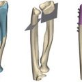
It is recommended that excision of the ulnar anlage be performed at age 6 months. Proponents of early excision state it may improve both the function and appearance of the arm [1, 7, 22]. The anlage acts as a tether and will restrict radial growth and increase deformity of the forearm. In addition, the forearm will double in length by age 3 years, and resection of the anlage will provide the best possibility for unrestricted growth of the limb [16, 22].
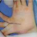
To excise the ulnar anlage, either a longitudinal or lazy “S” incision is used over the ulnar border of the forearm and wrist. Usually the flexor carpi ulnaris is absent and the neurovascular bundle (if present) is directly under the skin and needs to be protected. Distally, it is crucial to dissect the anlage off of the carpus and radius completely. Following distal resection, the wrist should be passively corrected to a neutral position. Resection of the entire fibrous anlage proximally is not required, and usually resection of the distal third is adequate [16]. If excessive bowing of the radius is present, then an osteotomy can be performed at the same time. Postoperative course includes immobilization of the wrist in a neutral position for 6 weeks followed by stretching and splinting for at least 6 months, although some authors have recommended nighttime splinting with a short arm orthosis until skeletal maturity [23].
Related posts:


Stay updated, free articles. Join our Telegram channel

Full access? Get Clinical Tree


