Chapter 16 Transverse upper body lift
• Laxity of the upper back is not corrected by the traditional lower body lift as the midline zone of adherence prevents tension from being transmitted to the upper back.
• The entire back deformity including skin laxity, excess adiposity and redundant lateral breast can be corrected using the Bra Line Back Lift.
• This technique is useful to contour the back in massive weight loss patients or in individuals showing signs of redundant skin and adiposity as a result of age.
• The scar is well-tolerated by patients and easily concealed beneath a bra or bathing suit top of the patient’s own choosing.
• The procedural learning curve is gentle for any plastic surgeon with experience in excisional body contouring.
Introduction
A number of methods have been utilized to remove and contour the excess tissue that can occur as a result of normal aging or after fluctuations in weight. Many of these techniques describe tissue removal along a dermatome pattern;1–15 however, this tends to leave an oblique scar that is difficult to conceal in normal clothing. Beginning in 2001, and first published in 2008, we developed a technique of transverse upper back lifting, described as the Bra Line Back Lift, to correct the deformity and place the final scar where it is easily concealed beneath a bra or bathing suit top.16 This procedure has been highly efficient in providing complete correction of skin laxity and excess adiposity in both the massive weight loss and normal weight population.
Preoperative Preparation
Patient Evaluation
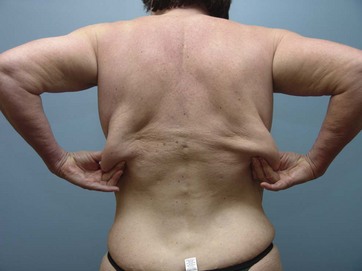
Patients with complaints of upper back tissue excess are evaluated for skin quality, stretch marks, subcutaneous adiposity and excess or hanging skin. Surgical judgement and patient wishes are used to formulate a surgical plan. Some patients are better candidates for standard tumescent liposuction, adjunctive liposuction procedures using laser or ultrasound, or direct excision in the form of a Bra Line Back Lift. This chaper will focus on excisional lifting (Fig. 16.1).
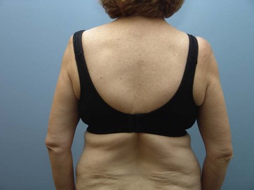
The patient is examined in a full-length mirror where they can confirm the areas in need of correction. Redundant tissue is firmly grasped and brought together using bimanual palpation to demonstrate to the patient what the final outcome will be in terms of resected tissue. Because the incision begins at the lateral inframammary fold, we also manually gather excess tissue from the lateral breast area to demonstrate the flank improvement which is often desirable to the patient. We ask patients to bring in their most revealing bra or bathing suit top so we can discuss scar placement and ensure their expectations will be met (Fig. 16.2). We also discuss the importance of keeping the arms abducted until full tissue relaxation has occurred to make sure the patient is comfortable with the post operative requirements of the procedure. Full range of motion is permitted by week six.
Preoperative Markings
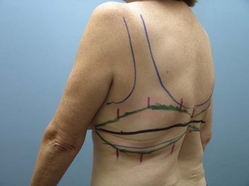
Markings are made in the standing position. We utilize a standard six position photographic technique to record the entire lateral and upper back condition. The six positions are left lateral, left oblique, posterior, right oblique, and right lateral. Boundaries of the patient’s ideal scar position are confirmed by marking the borders of a bra or bathing suit top of the patient’s choice. If the patient does not have a desired undergarment, the ideal final incision line placement is determined by identifying the level of the inframammary fold bilaterally and transcribing that across the back in a horizontal fashion (Fig. 16.3). When the final incision line has been determined, bimanual palpation is used to strongly gather the redundant skin and excess adiposity such that this redundancy centers the final incision line (Fig. 16.4). This gathering of tissue is performed at multiple spots across the entire line of resection and marked. These points are then connected as a continuum identifying the final area for resection. Note that the resection is the least in the midline because of the strong zone of adherence there (Fig. 16.5). It is this strong adherence along the spine that permits forces from lower body lifting from having any dramatic effect on the upper back. The resection is strongly tapered into the inframammary fold at the level of the anterior axillary line to avoid a dog ear. Realignment marks are then made across the area for resection to allow for precise realignment intraoperatively (Fig. 16.5). The markings are made with the arms at the patient’s side, allowing for the maximum amount of tissue to be gathered to achieve an optimal result.
< div class='tao-gold-member'>
Stay updated, free articles. Join our Telegram channel

Full access? Get Clinical Tree


