Fig. 40.1
Anatomy of the nerves of the mystacial pad. 1 zygomaticoorbital, 2 buccolabial, 3 upper marginal mandibular, 4 infraorbital
Our previous investigations showed the importance of facial nerve repair to achieve sensorimotor recovery in the hemifacial transplant model in rats [34]. The duration of the surgery and the high mortality rates were the main drawbacks of hemifacial transplantation. A modification to the hemifacial flap was developed to reduce the length of the procedure while preserving a functional unit of the rat face, which lead to a new flap design, properly named the mystacial pad flap.
The Experiment
A total of 56 animals were studied in six groups, as shown in Table 40.1. The “graft” term was assigned to those non-vascularized composite tissue transplants, while the “flap” term was used when vascular repairs were performed. In group I (anatomic study group, n = 8), Lewis rats were subject to harvesting technique trialing and the mystacial vascular network was drawn. In group II (flap viability group, n = 8), the mystacial pad elevated and reset in Lewis rats to check flap survival based on the facial vessels. In group III (isograft group, n = 8), viability without vascular pedicles was explored in Lewis to Lewis transplantation. In this group, a mystacial defect was created in the recipient, where the mystacial pad was set as a composite graft. In group IV (isoflap group, n = 8), vascularized mystacial pad flaps were transplanted from Lewis to Lewis rats and nerves were repaired, to serve as controls of the alloflaps. In group V (allograft group, n = 8), graft viability without vascular repairs was explored in Lewis-Brown-Norway to Lewis transplantation. Group VI was the study group, in which transplantations from Lewis-Brown-Norway to Lewis were done. Group VIa (non-neurotized alloflap transplant group, n = 8) represented the vascularized mystacial pad flap transplantation in the absence of nerve repairs, while in group VIb (neurotized alloflap transplant group, n = 8), the mystacial pad flaps were transplanted and the BL, uMM and ZyO branches of the facial nerve, and the Io nerve were repaired.
Table 40.1
Comparisons after mystacial pad allotransplantation
IV (n = 7) | VIa (n = 7) | VIb (n = 7) | pa | pb | pc | pd | ||||
|---|---|---|---|---|---|---|---|---|---|---|
Native whiskers | Transplanted whiskers | Native whiskers | Transplanted whiskers | Native whiskers | Transplanted whiskers | |||||
Conduction | ||||||||||
Duration | 1.40 ± 0.08 | 1.47 ± 0.03 | 1.30 ± 0.03 | 0 | 1.40 ± 0.03 | 1.47 ± 0.06 | 0.125 | 1.000 | 0.001 | 0.028 |
Amplitude | 5.06 ± 0.17 | 1.90 ± 0.16 | 4.52 ± 0.47 | 0 | 5.01 ± 0.13 | 1.93 ± 0.14 | 0.017 | 0.710 | 0.001 | 0.017 |
Resting needle (%) | ||||||||||
Electrical silence | 100 | 100 | 100 | 0 | 100 | 100 | – | – | 0.001 | – |
Denervation activity | 0 | 0 | 0 | 100 | 0 | 0 | ||||
Whisking needle (%) | ||||||||||
Normal | 100 | 0 | 100 | 0 | 100 | 0 | ||||
Moderate | 0 | 100 | 0 | 0 | 0 | 100 | – | – | 0.001 | – |
Denervation activity | 0 | 0 | 0 | 100 | 0 | 0 | ||||
Sensitivity test (%) | ||||||||||
Defense | 100 | 100 | 100 | 0 | 100 | 100 | – | – | 0.001 | – |
No response | 0 | 0 | 0 | 100 | 0 | 0 | ||||
Histology (%) | ||||||||||
Neurokeratin + | 100 | 100 | 100 | 0 | 100 | 100 | – | – | 0.001 | – |
Neurokeratin − | 0 | 0 | 0 | 100 | 0 | 0 | ||||
The Mystacial Pad Flap
The mystacial pad flap was elevated under microscope magnification (Leyca Microsystems, M300, magnification × 10 × 25 × 40). The skin was incised deep to the platysma in the anterior neck, and superficial to the facial muscles in the mystacial pad and frontal region. In the neck, the external jugular vein (EJV) and anterior facial vein were preserved, but the posterior facial vein was ligated. The submandibular gland was excised after ligation of the glandular branches of the facial artery and vein. The sternocleidomastoid muscle and facial vein were retracted to expose the CCA and its main branches, the external (ECA) and internal carotid arteries. The posterior belly of the digastricus muscle was excised, the omohyoid muscle was transected and the greater horn of the hyoid bone was excised. The hypoglossal nerve crossing anteriorly to the ECA needs to be transected. The facial artery is included in the flap as the main vessel to the mystacial pad. The internal carotid artery, superior thyroid artery, ascending pharyngeal artery, lingual artery, ascending palatine artery and the superficial temporal artery were ligated and transected. The facial artery and vein were undermined from the masseter muscle to the labial commissure, and protected creating a cuff of muscle to avoid skeletonization of the vessels. At this point uMM and BL branches were transected. The animal was placed laterally and the incision runs peri-oro-nasally. The posterior incision in the mystacial pad reached the frontal area to harvest the ZyO branch. From the frontal incision dissection was carried out downwards subperiostically and then just over the oral mucosa to include the facial vessels, reaching the insertion of the masseter muscle tendon. The fan IoN was transected proximally. The flap was elevated based on the anterior facial vein draining to EJV and on the facial artery off the ECA [35] (Fig. 40.2). The vascular pedicle was only transected at the end of the recipient preparation to minimize the time of warm ischemia.
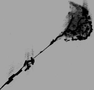
Fig. 40.2
Mystacial fat pad gelatin-barium roentgenogram showing vascular network of the flap
Preparation of the Recipient
The midline skin of the recipient’s neck was incised to approach the EJV and CCA. The sternocleidomastoid was freed from the sternoclavicular insertion and removed. The vagus and frenicus nerves were preserved. The incision ran to the labial commissure, where the facial artery was ligated, and then surrounded the mystacial area preserving the eyelids, nose and lips. The BL, uMM, ZyO branches were transected and the mystacial pad was removed leaving the oral mucosa intact. The IoN was transected as distal as possible for ease of repair. Then the mystacial pad flap was transferred to the recipient. End-to-end intimo-intimal EJV anastomosis was performed using 10-0 nylon sutures. Next, end-to-side CCA anastomosis was performed using 10-0 nylon. Then BL, uMM, and ZyO were sutured by epineural repair using 10-0, while the fan IoN was repaired with 8-0 (Figs. 40.3 and 40.4). Finally the skin was closed using 6-0 running absorbable suture. After the transplant the recipients received 2 mL of subcutaneous lactated Ringer’s solution to compensate for perioperative fluid loss.
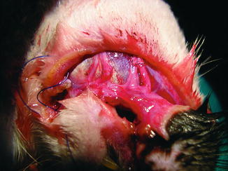
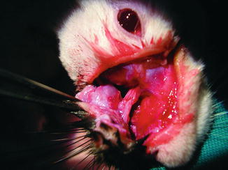
Fig. 40.3
End-to-end external jugular vein anastomosis, and end-to-side common carotid artery anastomosis on the recipient rat
Fig. 40.4
Epineural repair of the buccolabial and upper marginal mandibular branch of the trigeminal nerve and infraorbital nerve
Tapered subcutaneous tacrolimus (Astellas Pharma, Munich, Germany) immunosuppression monotherapy was initiated after surgery in groups V and VI. The tapering scheme consisted of 6 mg/kg/day during the first week, 4 mg/kg/day in the second week, and 2 mg/kg/day thereafter. The animals were weighted weekly for dosage preparation.
Anatomical, Clinical, Electrodiagnostic and Histological Studies
In group I, the vascular territory of the mystacial pad was drawn using radiographic studies by injecting a mixture of gelatin (market available) and barium (Barigraf, Juste, Madrid, Spain) into the common carotid artery (CCA) of two animals.
The general condition of the recipients in the other groups was observed daily for 8 weeks, and weight was measured every week. A clinical sign of graft rejection was defined as post-transplant hair loss, while hard touch was assumed to be a clinical sign of necrosis. Electrodiagnostic (EDX) evaluation was performed after 6 weeks under ketamine sedation (10 mg/kg) by a neurophysiologist unaware of the nerve repair procedures. Two percutaneous electrodes (one active, one reference) were placed at the retroauricular region to stimulate the facial nerve. A bipolar receptor electrode was placed percutaneously in the mystacial pad. The stimulation voltage was 2.8 mA, the electrical pulses were of 1 ms duration, and the frequency was 1 Hz. The presence, duration and amplitude of the conduction potentials were recorded if present in the conduction study. The presence of denervation activity or electrical silence during rest was noted. Motor unit potentials were also registered during whisking, and the records were classified into normal activity, moderate activity, or absence of activity (denervation activity) according to the neurophysiologist subjective criteria. After 8 weeks the animals were euthanized and biopsies of the mystacial region were taken. They were formalin-fixed and paraffin embedded; 5-mm thick sections were examined microscopically (Olympus Vanox AH-3, Japan) at ×300 and ×600 magnification after hematoxylin-eosin staining for the presence of the neurokeratin artifact. It traduces the presence of spirally rolled Schwann cells plasmatic membrane around the axons, providing the myelin to the nerve fibers. In the absence of axons, Schwann cells do not roll and the neurokeratin artifact is not seen. The presence of neurokeratin was noted for each sample as positive or negative by the first author, who was aware of the nerve repair procedure.
Statistical Analysis
Quantitative variables are presented as mean ± standard deviation and qualitative variables as percentages. Pairwise comparisons in the duration and amplitude of conduction studies in groups IV and VIb based on the explored side were performed using the Wilcoxon test. Mann- Whitney U test was used for the comparison of these variables in the transplanted side between IV and VIb and also between VIa and VIb groups. The differences in resting and whisking EDX, sensitivity test, and histology in the transplanted side based on these groups were analyzed using the Fisher exact test. Statistical significance was taken if P ≤ 0.05. Statistical analysis was performed using the SPSS Statistical Package, version 15 for Windows (SPSS Inc, Chicago, IL).
Results
All the results are summarized in Table 40.1. In group I the vascular axis of the mystacial pad, consisting of the angular branch and the superior labial artery off the facial artery was confirmed (Fig. 40.2). In group II all the flaps and animals survived. In groups III and V grafts necrosed 7.3 ± 0.6 days after transplantation, with no other complications. Before allograft transplantations, control isoflap transplantations were performed. Success was achieved in seven cases (87.5 %), and the recipients survived for 8 weeks (electively). In group VI, the procedure required an average of 3.53 ± 0.74 h and the average ischemia time was 1.13 ± 0.35 h. Fourteen recipients (87.5 %) showed perfect flap viability without signs of infection or rejection for 8 weeks (Fig. 40.5). Clinical explorations in group IV showed defense reactions both in normal and transplanted sides. In group VIa explorations showed no response when the whiskers of the transplanted mystacial pad were pulled, whereas defense reaction was seen in the native side. Explorations in group VIb showed defense reaction both in the native side and in the transplanted whiskers. Comparisons of the native side and the transplanted side in subgroup VIb revealed statistically significant differences in the duration and amplitude of the conduction potentials (P ≤ 0.028 and P ≤ 0.017, respectively).
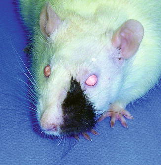
Fig. 40.5
Appearance of the recipient of a mystacial pad flap allotransplant after 8 weeks
The hematoxylin-eosin biopsies revealed the entrance of the nerve fascicles into the vibrissal follicles. Nerve sheaths in subgroup VIa did not show the neurokeratin artifact in any of the cases (Fig. 40.6). All biopsies in groups IV and VIb showed the neurokeratin artifact in the nerve sheaths (Fig. 40.6).
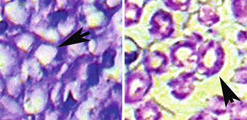
Fig. 40.6
Neurokeratin artifact in the nerve sheaths of groups IV and VIb
Discussion
The first scientific report of a FAT in humans was the case of a woman receiving a VCA consisting of nose, lips, chin and cheeks [3]. Despite the recipient was not free of immunologic rejection, a remarkable cosmetic and functional outcome was achieved [3, 36]. This excellent functional outcome was to be expected in a manner similar to hand and larynx transplants [4, 37]. Our investigations were conducted to clarify the return of motion and sensitivity to a functional subunit of the rat face after allotransplantation. The vibrissal trigeminal loop is an excitatory reflex arc that controls the mystacial musculature and depends both from its own level of activation and from the environment findings. Thus it can be studied independently of voluntary whisking [33]. We were able to demonstrate the return of sensitivity after neural repair in mystacial pad transplants in addition to histological and EDX signs of recovery.
Previous reports in hind-limb allotransplantation in rats had shown that sensory nerve function could be restored between 4 and 6 weeks, and motor function returned after 6–8 weeks. The recipients were able to stand on both extended hind limbs and climb vertically, with normal gait in 80 % [38–41]. Similarly, mystacial pad functional recovery was found in a period less than 8 weeks in the current work, and also in our previous report [34].
The path to develop the mystacial pad flap relied on previous face transplant research in rats (Table 40.2). Full face transplant model was described in rats for the first time in 2003 [24]. High mortality rates were seen until the model was established, surgeons’ skills improved and surgery length decreased. Harvesting the flap based on CCA and EJV bilaterally took about 2.5 h in the best hands. Further investigations led to creative vascular repairs to reduce vascular complications [19
 Microcirculation Model for Invasive Animal Monitoring
 Composite Osseomusculocutaneous Thymus Allotransplantation Model
 In Vivo Chimera Model: Creation of Primary and Secondary Chimera
 Peripheral Nerve Surgery Models Crush Injury and Epineural Patch
 Heterotopic Vascularized Ovarian Autotransplantation Model in the Sheep
 Defect Repairs After Resections of Laryngeal Cancer, Hypopharyngeal Cancer, and Cervical Esophageal Cancer


Related posts:
Microcirculation Model for Invasive Animal Monitoring
Composite Osseomusculocutaneous Thymus Allotransplantation Model
In Vivo Chimera Model: Creation of Primary and Secondary Chimera
Peripheral Nerve Surgery Models Crush Injury and Epineural Patch
Heterotopic Vascularized Ovarian Autotransplantation Model in the Sheep
Defect Repairs After Resections of Laryngeal Cancer, Hypopharyngeal Cancer, and Cervical Esophageal Cancer
Stay updated, free articles. Join our Telegram channel
Full access? Get Clinical Tree
