Fig. 64.1
View of the epineural sheath tube with the straight irrigator inside
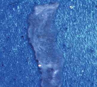
Fig. 64.2
View of the epineural sheath patch after splitting the epineural sheath tube
Application of Epineural Sheath Patch
Adult Lewis rats weighing between 250 and 300 g are used as recipient animals in this model. Preparation of the animal and sciatic nerve dissection is described at Chap. 64. After exposure of the sciatic nerve, approximately 2 cm above the trifurcation, about 4 mm nerve segment is marked and a circumferential epineurectomy is performed using microsurgical scissors under surgical microscope. After removal of the epinerium, the nerve fascicles are bulged. These epineurectomies are clinically relevant because a local epineurectomy is often performed during the course of nerve decompression surgery [2, 10]. Then the sciatic nerve segment that underwent epineurectomy, is crushed using 5-in. hemostat for 5 min as previously described in Chap. 64. After releasing the clamp, a loosely placed 10–0 nylon suture is tacked to the normal epinerium distally to guide the distal end of the epineurectomy. The harvested epineural sheath patch is wrapped around the crushed sciatic nerve with inner surface adjacent to the crushed nerve fascicles (Fig. 64.3). Then the epineural sheath is loosely fixed to the underlying muscle fascia with 10/0 nylon sutures to prevent migration of the epineural sheath patch. During implantation, nerve fascicles can easily be visualised through the semitranslucent epineural sheath patch that prevents additional travma to the nerve fascicles.
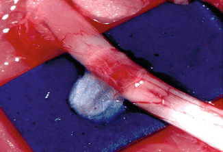
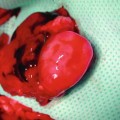
Fig. 64.3
Application of epineural sheath patch over crushed sciatic nerve segment
Before final closure, the muscles surrounding the sciatic nerve segment can be crushed using hemostats and/or be damaged using bipolar cautery to create an unfavorable wound condition that may mimics clinical case scenarios. Next, the gluteal muscles are reattached and skin is closed with interrupted absorbable sutures. The contralateral site is not operated on and is used as control. Postoperative care is described at previous chapter.
Evaluation Methods
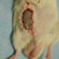
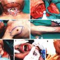
After functional and electrophysiological evaluations are done as described at previous chapter, the wrapped sciatic nerves are dissected for macroscopical assessment and histomorphological sampling. The epineural sheath patch could be identified under operating microscope and the absorption, degradation, adhesions, local inflammation, infection, hematoma, seroma and scar tissue formation can be observed. In the literature few adhesion scales are described for assessment of the adhesion quality, tenacity, extent of site involvement, and overall impression [1, 9]. Although these scales are semiquantative, they can still provide valuable clinical information and these assessments can be verified by the histological samples. The ability of the nerve to glide through the wrapped epineural sheath patch can be assessed (Fig. 64.4). Finally, the wrapped nerve was divided into segments such as proximal, mid and distal to the wrap and nerve samples were taken for histomorphologic evaluations.
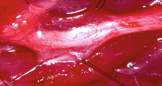
Fig. 64.4
View of the epineural sheath patch over the crushed sciatic nerve segment 6 weeks after the implantation. 9/0 nylon sutures are applied to the both sides of the epineural sheaath patch and pulled outwards to demonstrate the pliability of the patch
References
1.
2.
Related posts:
 Microcirculation Model for Invasive Animal Monitoring
 Composite Osseomusculocutaneous Thymus Allotransplantation Model
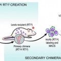 In Vivo Chimera Model: Creation of Primary and Secondary Chimera
 Experimental Model for Monitoring of Composite Tissue Transplantation Induced Trauma
 Heterotopic Vascularized Ovarian Autotransplantation Model in the Sheep
 Defect Repairs After Resections of Laryngeal Cancer, Hypopharyngeal Cancer, and Cervical Esophageal Cancer
Stay updated, free articles. Join our Telegram channel

Full access? Get Clinical Tree


