| Disfiguring proptosis |
| Compressive optic neuropathy unresponsive to maximally tolerated medical therapy |
| Exposure keratopathy unresponsive to maximally tolerated medical therapy |
| Preparation for strabismus surgery with large angle deviation |
| Deep aching pain and pressure from proptosis |
| Exophthalmometry readings |
| Extraocular muscle movements and primary deviation |
| MRD1, MRD2, degree of upper and lower eyelid retraction, lagophthalmos |
| Optic nerve function – relative afferent pupillary defect (RAPD), color perception, visual field testing, optic nerve head examination |
| CT scan – sinus relationships, muscle size and bone volume |
| Thyroid status including thyroid function tests |
| Area of decompression | Proptosis reduction |
|---|---|
| Orbital fat | 2 mm |
| Orbital fat and lateral wall | 4 mm |
| Orbital fat, lateral wall, and medial wall | 6 mm |
| Orbital fat, lateral wall, medial wall, and floor | 8 mm |
| Orbital fat, lateral wall, medial wall, floor, and lateral rim removal | 10 mm |
Introduction
Thyroid-related orbitopathy (TRO) is the most common cause of proptosis in adults. TRO is caused by an autoimmune process that leads to deposition of extracellular matrix in the orbital soft tissues that attracts water, causing swelling. This primarily occurs in the extraocular muscles; however, some patients have activation of orbital fat, leading to adipogenesis and fat hypertrophy. As with most autoimmune diseases, women have a 6 : 1 predilection for the disease. Factors associated with worsening disease include smoking, but tobacco use is not causal.
The most common eye manifestation is eyelid retraction, but other findings include: proptosis, restrictive strabismus, chemosis, conjunctival injection, eyelid edema, eyelid erythema, and optic neuropathy in severe cases. Approximately 90% of patients with eye findings are hyperthyroid, with the remaining 10% being either euthyroid or hypothyroid. CT imaging can reveal enlarged extraocular muscles (type II disease) and/or enlarged fat compartments (type I disease) in the orbit. The most commonly enlarged extraocular muscles in decreasing order are: inferior, medial, superior and lateral rectus muscles. The tendon insertions are typically spared from enlargement compared to myositis seen with idiopathic orbital inflammation.
Treatment is based on symptoms and severity of disease. Patients with optic neuropathy require urgent medical treatment followed by orbital decompression if resistant. In patients requiring elective surgical intervention, a staged approach is recommended (with not all stages being required) in order: orbital decompression, strabismus surgery, and eyelid surgery.
The decision to perform orbital decompression depends on many factors. The most common indication is disfiguring proptosis ( Table 64.1 ). The orbital aspect of the autoimmune process is self-limited and many patients have spontaneous improvement once the active inflammatory phase ends. Elective surgical rehabilitation should occur in the quiescent phase. Orbital decompression is an effective operation to help restore the proptosis to the pre-disease state. The preoperative evaluation must include measurement of exophthalmos and imaging ( Table 64.2 ). Symptoms of deep ache and pressure often resolve after orbital decompression. Our algorithm for decompression is seen in Table 64.3 .


Related posts:
 Mustardé’s epicanthoplasty
 Foundations of oculofacial plastic surgery
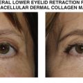 Lower eyelid retraction repair with porcine acellular dermal collagen matrix
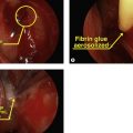 Endoscopic dacryocystorhinostomy with lacrimal sac biopsy
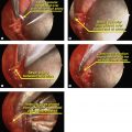 Transcaruncular approach to ethmoidal artery ligation
 Orbital implant exchange with dermis fat graft
Stay updated, free articles. Join our Telegram channel

Full access? Get Clinical Tree


