Thermal Injuries: Introduction
|
Epidemiology
The very young and very old are at increased risk of domestic burns.1,2 Active young adults in industrial jobs are at modest increased risk. In developed countries, about 70% of pediatric burns are caused by hot liquid. Flame injuries are more common in older children and young working adults. Scalding and flame injuries each account for approximately half of burn injuries in the elderly, with kitchen and bathing accidents being predominant. Approximately 20% of burns in younger children involve abuse or neglect.
Etiology and Pathogenesis
The development of an envelope of cornified skin was a crucial component of the adaptation of aquatic sea animals to the land environment. The vapor and fluid barrier created by the epidermal layer facilitates the maintenance of fluid and electrolyte homeostasis within very narrow limits. The dermis provides strength and flexibility, and the reactive dermal vasculature facilitates control of internal body temperature within very narrow limits. The appendages provide lubrication and prevent desiccation. All of these critical functions are lost when substantial areas of the skin are burned.
There is both a local and a systemic response to the burn wound.3,4 The local response consists of coagulation of tissue with progressive thrombosis of surrounding vessels in the zone of stasis over the first 12–48 postinjury hours. An ability to truncate this secondary microvascular injury and its associated tissue loss is a major area of ongoing investigation. In larger burns, a systemic response develops that is driven initially by release of mediators from the injured tissue, with a secondary diffuse loss of capillary integrity and accelerated transeschar fluid losses. This systemic response is subsequently fueled by by-products of bacterial overgrowth within the devitalized eschar.
Burn wounds are initially clean but are rapidly colonized by endogenous and exogenous bacteria.5 As bacteria multiply within the eschar over the days following injury, proteases result in eschar liquefaction and separation. This leaves a bed of granulation tissue or healing burn, depending on the depth of the original injury. In patients with large wounds involving 40% or more of the body surface, the infectious challenge generally cannot be localized by the immune system, leading to systemic infection. This explains the rare survival of patients managed in an expectant fashion with burns of this size.
The systemic response to injury is characterized clinically by fever, a hyperdynamic circulatory state, increased metabolic rate, and muscle catabolism.6 This stereotypical response to injury has been retained by all mammalian species. It is effected by a complex cascade of mediators, including changes in hypothalamic function resulting in increases in glucagon, cortisol, and catecholamine secretions; deficient gastrointestinal barrier function with translocation of bacteria and their by-products into the systemic circulation; bacterial contamination of the burn wound with systemic release of bacteria and bacterial by-products; and some element of enhanced heat loss via transeschar evaporation. It is likely that this response has significant survival value, but control of some of the adverse aspects of this response, particularly muscle catabolism, is an active area of ongoing investigation.7,8
Clinical Findings
|
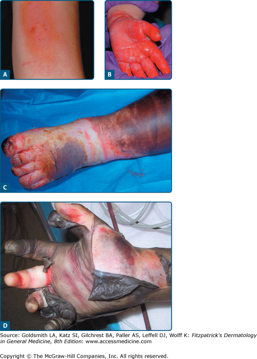
- First degree: Red, dry, and painful and are often deeper than they appear, sloughing the next day (Fig. 95-1A).
- Second degree: Red, wet, and very painful with enormous variability in their depth, ability to heal, and propensity to hypertrophic scar formation (see Fig. 95-1B).
- Third degree: Leathery, dry, insensate, and waxy (see Fig. 95-1C).
- Fourth degree: Involve underlying subcutaneous tissue, tendon, or bone (see Fig. 95-1D).
Figure 95-1
Burn depth is classified as first, second, third, or fourth degree. A. First-degree burns are red, dry and painful and are often deeper than they appear, sloughing the next day. B. Second-degree burns are red, wet and very painful. There is an enormous variability in their depth, ability to heal, and propensity to hypertrophic scar formation. C. Third-degree burns are leathery, dry, insensate, and waxy. D. Fourth-degree burns involve underlying bone and/or muscle.
Lightning Strikes
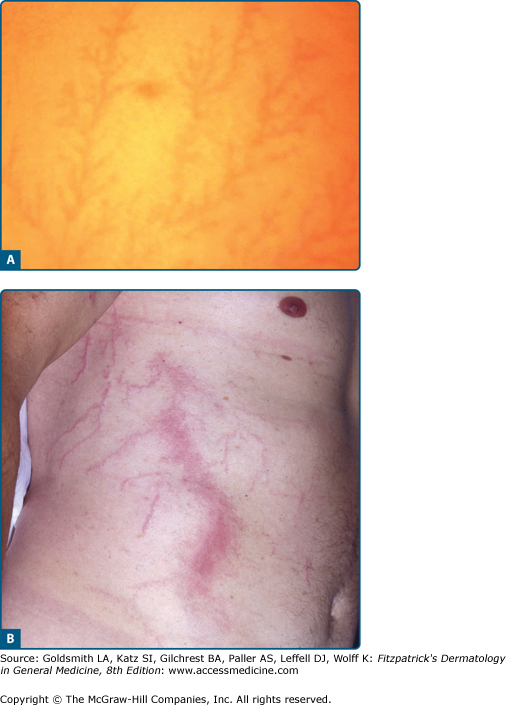
Although lightning strikes carry large amounts of energy, measured in millions of volts, human injuries are rare, with only about 50 people so injured annually in the United States and a fatality rate of only about 10%. Injury can rarely occur from direct strike, and is usually fatal. More commonly, people are injured by current flow around the skin envelope, or side flash, when a nearby object is struck. Destructive burns are unusual. Serious injury is more often the result of associated blunt trauma or occasional cardiac arrest. In most survivors, lingering symptoms are nonfocal, neurologic in nature, often preceded by an immediate but transient loss of consciousness.9 The most common physical finding in those injured by side flash is an evanescent serpiginous cutaneous erythema, as noted in Figure 95-2.
Complications
In the outpatient setting, patient selection should ensure that major complications are few (Table 95-1). The most common issues that arise are wound sepsis, excessive pain and anxiety, and underestimation of burn depth.10 The most common wound infection in this setting is streptococcal cellulitis, which presents initially with surrounding erythema that progresses to lymphangitis and systemic toxicity (Fig. 95-3). These patients often need admission for antibiotics, observation, and sometimes surgery. In some situations, adequate pain and anxiety management is very difficult in the outpatient setting, especially around dressing changes. This can be addressed with judicious medication and sometimes carefully monitored membrane dressings. Some patients will require admission. It is common for burn depth to be underestimated initially, with areas of full-thickness injury not appreciated for several days. These patients may require admission for surgery.
Common Outpatient Burn Complications Stay updated, free articles. Join our Telegram channel
Full access? Get Clinical Tree
 Get Clinical Tree app for offline access

|


