The propeller flap, based on a single vascular pedicle supplying a fasciocutaneous island of skin, is a very useful technique to reconstruct soft tissue defects and has wide applications throughout the body. The use of this unique flap is pushing the boundaries of local flap reconstruction and bringing up intriguing questions about our understanding of the vascular basis of fasciocutaneous flaps.
Evolution
Wounds on the distal third of the lower extremity are known to be difficult to reconstruct. The lack of spare local tissue in the immediate vicinity of such wounds makes it difficult to design local flaps. With unreliable outcomes and increased morbidity, the general perception is that a free flap is the preferred option. However, I have always found it difficult to accept that a small to medium-sized defect on the distal third of the lower leg can only be reliably covered by a free flap.
In the 1980s, Ponten and Barclay and colleagues published the first clinical reports on fasciocutaneous flaps showing that long flaps can safely stretch the length to width ratio of random pattern flaps from 1:1 to 3:1. Their results generated much interest and stimulated many studies into peripheral vascularization of the skin through perforators. Surgeons who realized the importance of perforators began to preserve the myocutaneous or septocutaneous perforators when designing new flaps. Donski and Fogdestam demonstrated that such long flaps could just as safely be based distally on the lower peroneal perforators and used for reconstruction of defects around the lower third of the leg. These investigators postulated that by keeping the long axis of the flap on top of the posterior peroneal intermuscular septum, the perforators linked within the flap, forming an “axial pattern” blood flow that was therefore able to sustain a longer flap than would otherwise be possible. Amarante and colleagues showed that the lower posterior tibial artery perforators could equally be used safely to base such peninsular fasciocutaneous flaps for covering defects around the lower third of the leg. Gradually, the design of the skin bridge at the base of such peninsular flaps was being made progressively narrower until some surgeons felt brave enough to totally divide it and completely island the flaps. These investigators realized that keeping the skin bridge at the base of a peninsular flap does not improve its blood supply and may in fact kink the pedicle, contributing to vascular embarrassment of the flap. Furthermore, it was evident that the bulky “dog ear” was visually unappealing. These surgeons were also the first to routinely visualize the perforators and eventually isolate the flap on to a single perforator, but they remained reluctant to handle or clean around the pedicle. Most reports were of very few cases until Erdmann and colleagues reported the first big series from which they concluded that the distally based islanded fasciocutaneous flap was a good first choice for coverage of defects on the lower third of the leg and ankle.
What is the propeller flap?
The propeller flap is a local island fasciocutaneous flap based on a single dissected perforator. It is designed like a propeller with 2 blades of unequal length with the perforator forming the pivot point so that when the blades are switched, the long arm comfortably fills in the defect ( Fig. 1 ). The ability of this flap to rotate any angle up to 180° makes it extremely versatile for reconstructing traumatic as well as other defects of the distal lower limb where it was originally conceived. Gradually its use has been extended for reconstruction of many defects of varying etiology throughout the body. The propeller flap concept is best explained by addressing 3 key questions.
Why Does it have to be Based Distally for the Distal Lower Limb Reconstruction?
The lower leg is shaped like a cone tapering down toward the lower third and ankle with a paucity of spare tissue in that area for use in reconstructing defects. For this reason a proximally based peninsular fasciocutaneous flap tends to struggle in terms of getting enough healthy tissue into the defect and, in addition, it risks exposing either the subcutaneous border of the tibia or the Achilles tendon, both of which are difficult to graft and could often be prone to unstable scarring in the long term. The propeller flap, pivoted on a single perforator, avoids these problems by importing truly undamaged tissue from the proximal calf into the primary defect. In doing so it simultaneously transfers the secondary defect to an easily graftable area over the proximal muscle bellies. Even better, when there is tissue laxity in the proximal calf, the propeller flap can often allow the secondary defect to be closed primarily.
Why Does it have to be Based on only a Single Pedicle?
A logical question, because if one perforator is safe surely two should be safer. However, the design of this flap makes the use of more than one pedicle a potential hazard because the two pedicles could kink each other ( Fig. 2 ) or, in trying to avoid this problem, the rotation of the flap becomes limited. When the rotation is needed only up to 90° it may not matter if more than one pedicle is kept. However, when the flap is needed to rotate 180° it is actually safer to divide all perforators except one.
Why Island the Flap?
This is also a safety issue because the skin connection of a peninsular flap makes an awkward and unsightly twist at its base, which could risk compressing and stretching the pedicle and potentially cause the flap to suffer. In contrast, the cutting of all the soft tissue connections to truly island the propeller flap gives it a much greater freedom to pivot and rotate around its pedicle. This procedure also allows for a more distal reach of the flap on to the dorsum and lateral aspect of the foot when needed. It is also easier to inset and gives a better final contour.
Surgical technique
Propeller Flap for Distal Lower Limb Defects
Guidelines
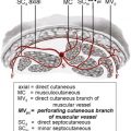
In the lower limb, the main useful perforators arise from the 3 major vessels—the posterior tibial, the peroneal, and the anterior tibial arteries. In my experience, it is easier to base the flap on the perforators arising from the first 2 vessels. As mentioned before, in designing the flap it is best to avoid transgressing onto the subcutaneous border of the tibia as well as the Achilles tendon. Also, it would be desirable to try to avoid damage to the saphenous nerve or the sural nerve depending on which side of the leg the flap is based.
It is important to adopt a flexible approach in the design and execution of the procedure, and although this may sound daunting to the beginner, following a few simple rules makes it a straightforward operation.
Design of the flap
Preoperatively, a handheld 8- to 10-MHz Doppler ultrasound scanner is helpful to locate the most promising perforator artery near the defect. A provisional flap design can then be drawn as follows, with the perforator as the pivot point of the flap ( Fig. 3 A). First, the distance between the perforator and the distal edge of the defect is measured. This value is then transposed proximally along the axis of the main source vessel, again measured from the perforator, and 1 cm is added. This value forms the proximal limit of the flap. Next, the width of the proximal flap needed to cover the defect is determined by measuring the width of the defect. This value is then used to determine the proximal flap width, adding 0.5 cm to allow for flap contraction and to facilitate its inset without tension. It is also important to ensure that, at the pivot point where the perforator pedicle enters the flap, the lateral dimensions are equidistant to ensure that when the flap is eventually rotated around to fill the defect there is no excessive sideways traction on the perforator during wound closure.
Related posts:


Stay updated, free articles. Join our Telegram channel

Full access? Get Clinical Tree


