This article reviews historical aspects of flap development, leading up to the exciting recognition of perforator flaps. The role and use of perforator-type flaps in the reconstructive armamentarium is reviewed as it pertains to different regions of the body.
The reconstructive surgeon needs to be familiar with the full spectrum of reconstructive options, and select the method of reconstruction that will give the best aesthetic and functional outcome. Perforator flaps are the most recent, and without doubt the most exciting addition to our armamentarium as reconstructive surgeons. New flaps are being described with great rapidity, and it can be difficult to keep up with the pace of progress occurring in our specialty.
This fact is especially true for surgeons who have been in practice for several years and who have already incorporated into their practice the many innovations that preceded popularization of perforator flaps. While it is important to keep up to date and be progressive, it is also important not to give up the true and tried methods that have been successfully used for years, thus risking doing harm to patients by attempting newly described procedures without adequate preparation. One has to be aware that every new procedure has a learning curve, which can be steep. Perforator flaps certainly fall into this category for the novice who is not experienced in the relevant anatomy and the requisite fine dissections. Furthermore, it is important to realize that not all new procedures will stand the test of time. Although they may progress through a period of popularity initially, any increased complication rate may relegate them to the “list of procedures that I no longer do,” a common list among some plastic surgeons.
As it turns out, many of the flaps that surgeons have used for years are in fact perforator flaps, although they have been known by another name. Without doubt, the recent emphasis on perforator flaps has enhanced our understanding of how flaps receive their blood supply. When it comes to raising flaps, it is the blood supply that is critical rather than the type of tissue in the flap. Thus, the change in nomenclature emphasizing the blood supply to the flap is a step forward. This advance is especially true for the musculocutaneous flaps, where the nutrient blood flow to the skin is dependent on perforating vessels and not the underlying muscle. There is no doubt that perforator flaps are here to stay and they have greatly increased the number of flap options for reconstruction. However, the extent to which the individual surgeon will incorporate newer perforator flaps into his or her “reconstructive ladder” or “elevator” will differ and continue to evolve. Ongoing debates and discussions on this exciting addition will be beneficial for our specialty.
Here the authors propose to briefly review the historical aspects of flap development and provide an overview of the senior author’s (J.J.P.) personal experience with perforator flaps. Further, this expansive choice of flaps has led to a change in emphasis in our reconstructive efforts, with improved selection and refinements.
A historical perspective
As already alluded to, the options available to the reconstructive surgeon have increased greatly over the last 40 years. It has been both exciting and a privilege to be a part of this process. It is hard to imagine what it was like to practice our specialty in the days of Sir Harold Gillies and other pioneers in the early part of the twentieth century, who were confronted with the daunting task of repairing terrible and devastating war injuries. Although these early pioneers were innovative, patient, and persistent, they had very few choices to draw upon. Gillies recognized that “tissue transfer was a constant battle between blood supply and beauty,” but it was the lack of detailed knowledge of the way skin obtains its blood supply that delayed advances at that time. Small defects were repaired with skin grafts or local random pedicle flaps, which were sometimes extended by applying the principles of delay. Other than the deltopectoral flap described by Bakamjian, there were very few regional flaps. For larger defects, multistaged tube pedicle flaps, first described by Filatov and popularized by Gillies and others, were used. This practice continued well into the 1960s.
The concept of axial flaps evolved in the 1970s, and a major discovery was the pedicle groin flap described by MacGregor and Jackson. A new era began when it was realized that axial flaps could be detached and transferred as free flaps. It was the development of the practice of microsurgery by Buncke and others that fueled the discovery of new flaps. The breakthrough occurred when the first cutaneous free flap was reported in Melbourne by Taylor and Daniel, soon followed by O’Brien and Shanmugan and several others. However, at that time there really were no other commonly known cutaneous flaps other than the groin flap, and the focus was on flap survival.
It became apparent that a more detailed anatomy of the vascular supply to the skin and its contents was required to not only increase the donor options, but to allow better flap selection and refinement. The outcome was a major renaissance in anatomic research. It was realized that in many regions of the body, the skin obtains its blood supply through the underlying muscles. This finding led to the discovery of many musculocutaneous flaps by Ger, Orticochea, McGraw, and others, thus greatly expanding the repertoire of the reconstructive surgeon.
At the same time Ponten observed that the inclusion of the deep fascia in extremity reconstruction led to hardier, more reliable flaps. These “super” flaps marked the beginning of fasciocutaneous flaps. As new muscle, musculocutaneous flaps, and fasciocutaneous flaps were being described, there was a real need to return to the cadaver laboratory to practice and perfect the harvest of these various flaps. Flap courses became common and an essential part of residency training. There followed an intense attempt to classify and categorize these newly described muscle and musculocutaneous flaps by Mathes and Nahai, and fasciocutaneous flaps by Cormack and Lamberty, among others. The reconstructive ladder was rapidly lengthening, with more options available. The focus then changed to flap refinement.
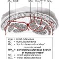
The body of knowledge was further augmented by Taylor and Palmer, who not only rediscovered the largely forgotten works of Manchot and Salmon, but also performed their own extensive injection studies and cadaver dissections. These investigators further elucidated the arterial, venous, and lymphatic circulation to the skin and its contents, enabling Taylor to propose the “angiosome concept” of interconnecting vascularity of the skin, which greatly helped our understanding of how flaps survive. The realization that the major source vessels supplied nutritional blood flow to all tissues to which they traversed, although obvious now, has not always been appreciated. These major vessels and their main named branches send other smaller branches to supply the surrounding muscles, bones, joints, and all connective tissue, and eventually the skin. It was discovered that in most locations there were set patterns in the way that skin obtained its blood flow: (1) via direct branches from source vessels that penetrated the fascia, (2) via vessels that first traversed muscle or passed through septa between the muscle groups, or occasionally (3) via vessels that first supply the bone, tendon, nerves, and so forth, giving rise to the concept of perforator flaps. Koshima, a superb ultra-microsurgeon, is credited with the early work that led to the practical application of perforator flaps.
Taylor’s anatomic dissections identified almost 400 good-sized arterial perforators (at least 0.5 mm diameter), which each supplied a volume of skin, and therefore were capable of serving as pedicles for flaps and theoretically could be detached and transferred as free flaps ; this obviously greatly increases the number of flaps available for reconstruction, and Wei and colleagues coined the term “freestyle flaps” to describe the many potential flaps that could be raised on any of these yet unnamed perforating pedicles. However, as more flaps were being reported, it again became necessary to categorize and classify these new modifications. A shift in emphasis from naming flaps according to the body of tissue it contained to its source blood supply was a major step forward. This change in nomenclature led to a better understanding and reflected a more profound awareness of why some flaps were not always reliable. An example of this is the gracilis musculocutaneous flap, which traditionally is raised with a vertical skin island overlying the muscle. It is well known that the distal part of this vertical skin island is not reliable.
Yousif and colleagues first pointed out that a transversely oriented skin island across the proximal third of the muscle is more reliable because it is richly supplied by the perforators from the medial circumflex femoral vessels that send branches through the muscle, as well as septal cutaneous perforators on either side of the muscle. Astute observations such as these help refine our reconstructive ladder.
With the potential for hundreds of new flaps, it became imperative to come to a consensus with naming and classification. An international meeting in Gent was helpful in developing the new nomenclature. There have also been several proposals for flap classification. Nakajima and colleagues have provided a very useful classification of the 6 different pathways through which blood flows from the source vessels to the skin. This classification has been further simplified according to the ease of pedicle dissection, as “direct perforators” from source vessel regardless of how it gets to the skin, and “indirect perforators” that are more difficult to dissect as these traverse the muscle first.
Related posts:
 The Anatomic Basis of Perforator Flaps
 Preoperative Imaging Techniques for Perforator Selection in Abdomen-Based Microsurgical Breast Reconstruction
 The Integration of Muscle Perforator Flaps into a Community-Based Private Practice
Perforator Flaps in the Upper Extremity
Pedicled Perforator Flaps in the Trunk
The Integration of Muscle Perforator Flaps into a Community-Based Private Practice
Perforator Flaps in the Upper Extremity
Pedicled Perforator Flaps in the Trunk
 Perforator Flaps in Breast Reconstruction
Stay updated, free articles. Join our Telegram channel

Full access? Get Clinical Tree


