The recent enthusiasm for perforator flaps underlines the need for a detailed understanding of the cutaneous vasculature. The principle determinant of success in perforator flap surgery is the inclusion of an adequately sized cutaneous perforator in the flap. Therefore, the size, distribution, and variability of cutaneous perforators of the human body are crucial to the design and execution of successful perforator flap surgery. Based on numerous anatomic studies, the authors have found that the main source arteries supplying the skin are fairly constant but the individual cutaneous perforators are quite variable. Knowledge of the overall architecture of the vasculature and an awareness of the variability, combined with a flexible operative plan, will enable the perforator flap surgeon to take advantage of the most appropriate perforators to execute a successful operative plan.
The vascular anatomy of cutaneous perforators is of vital importance for the design of successful perforator flaps. The detailed knowledge of this vascular anatomy provides the framework for flap elevation. Therefore, it is critical that the reconstructive surgeon has a detailed understanding of the vascular anatomy of the human integument. The purpose of this article is to provide an overview of the vascular anatomy of the human body to allow surgeons to use and customize perforator flaps in reconstructive plastic surgery.
Historical perspective
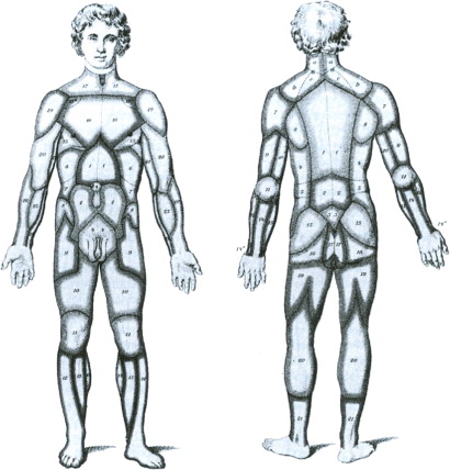
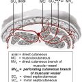
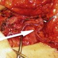
Harvey, in the 1600s, and Thomas and Spalteholz, in the 1800s, provided some of the earliest descriptions of the cutaneous vasculature. In the late 1880s, Carl Manchot performed a detailed study of the human cutaneous blood supply ( Fig. 1 ) and provided an early description of the vascular territories of the human body. This work was done by dissection alone, which is a remarkable feat given the complexity of the human skin vasculature. Manchot’s description of the cutaneous vasculature, and in particular the vascular territories of the integument, has stood the test of time. In the 1930s, Michel Salmon did a series of anatomic dissections using the original lead oxide injection technique and provided the earliest and detailed descriptions of the vascular supply to the skin ( Fig. 2 ). He contributed important concepts regarding the vasculature that remain relevant to the reconstructive surgeon, including observations about the pattern of cutaneous perforators. Subsequently, Ian Taylor and Palmer have provided comprehensive vascular studies of the human integument and together with John Palmer described the angiosomes of the body ( Fig. 3 ). The angiosome concept provided an important early framework for the development of perforator flaps. The use of the Doppler was described to identify the variable skin perforators. More recently, anatomic techniques have included a combination of vascular injection studies and three-dimensional (3D) computerized angiography to provide the most detailed illustrations of the vascular anatomy of the integument. The lead oxide injection technique was initially described by Michel Salmon in the 1930s. Rees and Taylor modified the technique and used it extensively in a series of anatomic projects. The authors have used this technique to comprehensively document the human cutaneous perforators.
Related posts:
 Where do Perforator Flaps Fit in our Armamentarium?
Preoperative Imaging Techniques for Perforator Selection in Abdomen-Based Microsurgical Breast Reconstruction
Where do Perforator Flaps Fit in our Armamentarium?
Preoperative Imaging Techniques for Perforator Selection in Abdomen-Based Microsurgical Breast Reconstruction
 The Integration of Muscle Perforator Flaps into a Community-Based Private Practice
Perforator Flaps in the Upper Extremity
Pedicled Perforator Flaps in the Trunk
The Integration of Muscle Perforator Flaps into a Community-Based Private Practice
Perforator Flaps in the Upper Extremity
Pedicled Perforator Flaps in the Trunk
 Perforator Flaps in Breast Reconstruction
Stay updated, free articles. Join our Telegram channel

Full access? Get Clinical Tree


