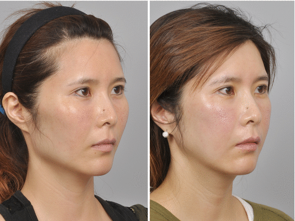
Fig. 18.1
Preoperative (left) and postoperative (right) views after conventional reduction malarplasty. Note the lack of reduction at the anterolateral malar protrusion, especially at the inferolateral orbital rim area after conventional reduction malarplasty procedure
Patient Assessment and Consultation
Patients who had previous conventional reduction malarplasty but were dissatisfied with the results seek secondary operation for further improvement as the zygomatic body and the inferolateral orbital rim continued to appear prominent. Some patients who complain of prominent cheekbones, which were assessed as difficult to be reduced by using conventional methods, are also proper candidates of orbito-malar complex reduction surgeries. All patients are analyzed by physical examination, along with clinical photographs and radiographic studies. Zygomatic arch view and three-dimensional computed tomography images are used for radiographic inspection to assess the extent of change before and after surgery. All patients have a detailed consultation with the surgeon to discuss their requirements and to understand the practical goals of the procedure before surgery.
Surgical Techniques
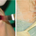
All patients are given general anesthesia. Orotracheal intubation is preferred at ID hospital, but nasotracheal intubation can be used. Intraoral, preauricular and subciliary, or transconjunctival approaches were universally applied. Intraoral and preauricular incisions are equal to those of conventional reduction malarplasty described at the previous chapter. Through these incisions, the soft tissues are elevated superiorly and laterally at the subperiosteal plane to expose the anteroinferior portion of the maxilla, zygomatic body, infraorbital foramen, and zygomatic arch with care taken to prevent infraorbital nerve injury. In contrast with conventional reduction malarplasty, dissection aims at exposing the zygomaticomaxillary suture as the osteotomy is performed along this suture line. For the subciliary approach, a skin incision is made inferior to the lower eyelashes, almost along the full length of the eyelid. To reach the orbital rim, a skin-muscle flap is elevated inferior to the lower eyelid. Preseptal transconjunctival approach is also useful especially in patients favoring invisible scars as possible. Lateral canthotomy can be considered to minimize the risk of periorbital tissue injuries and secure lateral orbital rim exposure. After the periosteum is incised, subperiosteal dissection was performed to expose the lateral and inferior aspect of the orbital rim. The extent of dissection of the lateral orbital rim is narrower than in conventional tripod osteotomy surgery, and the usual exposure of the zygomaticofrontal suture is not mandatory as the level of osteotomy and burring is inferior to the location of the suture.
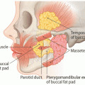
Tripod Osteotomy
In patients with moderate to severe orbital rim protrusion, tripod osteotomy is a more effective strategy. The orbito-malar complex is osteotomized at the zygomatic arch, zygomaticomaxillary suture, and about 5 mm inferior to the usual zygomatico frontal suture by a reciprocating saw and osteotome (Fig. 18.2). The walls of the lateral and inferolateral portion of orbit are osteotomized at about 5 mm from the margin of the orbital rim, on the inner aspect of the orbital walls. Osteotomy lines are designed as shown in Fig. 18.3. The application of the osteotome around the orbital rim requires gentle osteotomy and manipulation to avoid the risk of eyeball injuries, enophthalmos, and alteration in orbital volume. After the osteotomies are completed along the designed lines, the intervening bony segment is removed, shaved, and repositioned posteromedially as planned. The repositioning of the bony segment involved fixing with microplates and screws at the lateral orbital rim, inferior orbital rim, and zygomatic arch. Sophisticated burring is applied to palpable bony steps, if any, to obtain a smoother and more natural contour. Wound closure was performed after confirming bilateral symmetry.
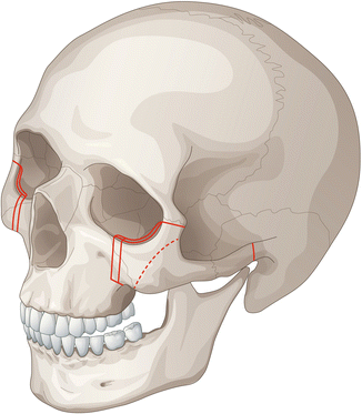
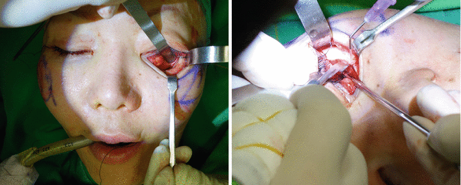
Fig. 18.2
Osteotomy lines of orbito-zygomatic reduction
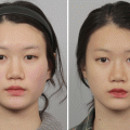
Fig. 18.3
Intraoperative photos. Osteotomy design of the orbital rim (left) and osteotomy using reciprocating saw and osteotome (right)
Orbital Rim Shaving
In patients with mild orbital rim protrusion or localized protrusion, the orbital rim can be shaved off by burring method. After completion of osteotomy, the channel retractor is inserted to protect the soft tissue around the orbit and burring the rim ends leaving enough the cortical bone to fix the plate with resected zygomatic body. Irrigation of areas leaving no bony dust is critical and let the soft tissue re-drape naturally around the region. Quite extensive burring of the orbital rim is possible and proves to be a very effective and versatile method for orbito-zygomatic prominence.
Key Technical Points
Conventionally, there are two surgical methods described for improving the prominent inferolateral orbital rim: one, shaving the orbital rim with a burr and, the other, mobilizing the orbital rim by tripod osteotomy. The shaving method is less invasive, but the rasping and burring of the orbital rim may be traumatic to the periorbital soft tissue, and the degree of improvement achieved may be inadequate. The tripod osteotomy, though more invasive, is considered to be more effective and reliable in achieving the desired improvement. Surgeons should make a decision which method is effective and efficient.
Case Study
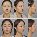
Case 1
A 34-year-old woman complained of prominent zygoma and wide midface (Fig. 18.4, left). We recommended orbito-zygomatic reduction for this patient as she had concurrent protrusion of her orbital rim and zygomatic body and arch. Osteotomy was performed to achieve a 5-mm reduction of each zygoma in order to reduce the protrusion of her zygoma. The posterior part of the zygomatic arch was divided by performing a complete osteotomy. The osteotomized orbito-malar complex was shifted medially (by 5 mm) and posteriorly (by 3 mm) and fixed by using microplates and screws. The prominence of orbital rim and zygoma markedly improved as seen at 6 months postoperatively (Fig. 18.4, right).