Fig. 19.1
Illustration of the mini-zygoma reduction surgery. First, osteotomy of the zygomatic arch is performed through a sideburn incision using a reciprocating saw. Then, osteotomy on the zygomatic body is done through a temporal incision with a J-shaped reciprocating saw
Fig. 19.2
Intraoperative photographs of the mini-zygoma reduction surgery. The zygomatic arch is completely osteotomized through a sideburn incision (above). The zygomatic body is osteotomized through a temporal incision, while the anterior side of the periosteum of the zygomatic body is left intact to avoid bone displacement (below). After all osteotomies are completed, the zygomatic arch is rigidly fixed with metal fixtures via the sideburn incision
After the osteotomy is completed on the zygomatic body, movability of zygomatic arch is visible through the sideburn approach. After all osteotomy procedures are completed, the arch is repositioned inwardly into the proposed position according to the preoperative surgical plan. Bone shaving by a surgical bur on the edge of the osteotomy margin may be performed to avoid palpability of the bony steps. After blunting the sharp osteotomy margins, the zygomatic arch is rigidly fixed using a prebent three-hole linear titanium plate and screws [3]. This allows the osteotomized arch segment to resist against a downward and rotational force which is mainly caused by the attached musculature (Fig. 19.3) [1].
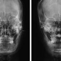
Fig. 19.3
Comparison on preoperative and postoperative imaging studies. Preoperative and postoperative three-dimensional computed tomographic images (above). Preoperative and postoperative zygomatic arch view radiographs (below)
Key Technical Points
- 1.
During the approach through the sideburn incision, blunt dissection for the subcutaneous fatty layer is recommended rather than the use of electrocautery or sharp scissors to avoid injury on the frontal branch of the facial nerve.
- 2.
Although the osteotomy is completed on the zygomatic body, it is important to confirm the mobility of the zygomatic arch before removing the saw from the zygomatic body osteotomy site because it is difficult for the surgeon to place the saw exactly on the previous osteotomy site if an additional sawing is required [1].
- 3.
In cases which the zygomatic arch protrusion starts behind the anterior tubercle of the zygomatic arch, additional burring on the posterior portion of the osteotomy site on the arch will be helpful in achieving a more favorable result [14].
- 4.
Patients may be discharged on the same day of surgery and informed to have soft diet for 2 weeks postoperatively. Trauma or direct pressure on the surgical site should be avoided for about a 6-week period after the surgery.
Case Study
Case 1
A 28-year-old man with an outward protrusion of the zygomatic arch underwent the mini-zygoma reduction surgery. A concomitant mandibular angloplasty was performed. The protrusion of the zygomatic bone is efficiently improved resulting in a reduction of the midface width and a smooth facial contour (Fig. 19.4).
Fig. 19.4
Preoperative view of a 28-year-old man who underwent the mini-zygoma reduction surgery (left) and postoperative view at 4 months after the surgery (right)
Case 2
A 21-year-old woman with lateral prominence in her midface region was planned for the mini-zygoma reduction surgery. Botulinum toxin injection was done together to treat her masseteric hypertrophy. The outwardly protruding portion of the zygomatic bone was reduced to result in a slender and smooth facial contour and to a more feminine appearance (Fig. 19.5).
Fig. 19.5
Preoperative view of a 21-year-old woman who underwent the mini-zygoma reduction surgery (left) and postoperative view at 3 months after the surgery (right)
Complications and Management
According to a previous study reported from the authors’ institution, the mini-zygoma reduction surgery was all performed under outpatient basis without any notable immediate complications such as unexpected bleeding or any degree of nerve injuries [1]. None of the patients required hospitalization for postoperative management. Other complications such as facial nerve paralysis, hematoma, infection, nonunion, or malunion of the bone segments were not reported throughout the same study.
Related posts:

Stay updated, free articles. Join our Telegram channel

Full access? Get Clinical Tree


