Fig. 20.1
Paranasal augmentation. Note that intraoral incision is made about 1 cm above the upper gingivolabial sulcus. Subperiosteal dissection exposes the area to be augmented. The infraorbital nerve should be preserved. Two points fixation with screws are recommended to prevent rotation of implant. The root of the canine should be avoided when the implant is immobilized with screws

Fig. 20.2
Implants for paranasal augmentation. The implants used in ID hospital are shown. It is made of silicon, triangular shape with concave medial side to fit to pyriform aperture. The thickness of implants ranges from 2 to 6 mm usually
Fig. 20.3
Suborbital augmentation. (a) Note that the infraorbital nerve should be identified and preserved during the subperiosteal dissection. To avoid irritation or compression to the infraorbital nerve, the superior border of the implant should be trimmed. The posterior surface of implant should be carved to obliterate the dead space above the anterior surface of the zygoma. Implants are fixed with titanium screws in two points for immobilization. (b) Implants may be positioned on zygomatic body to augment the maximum malar projection
Fig. 20.4
Implants for suborbital augmentation. The implants used in ID hospital are shown. It is also made of silicon like paranasal implants. Unlike the paranasal implants, the shape is ovoid to rectangular with round border. The thickness of implants ranges from 2 to 6 mm
Key Technical Points
- 1.
Proper selection of implant and placement are cornerstones in success.
- 2.
Dissection should be minimized but not too small to distort the implants.
- 3.
Immobilization with two points screws fixation give stable positioning of the implants.
- 4.
Clean operative field and bleeding control are important to prevent infection.
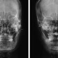
Case Study
Case 1
A 25-year-old man was planned for the malar reduction and V line surgery. But, he had depressed paranasal area because of recessed pyriform aperture. Thus, paranasal augmentation is performed using silicon implants of 5 mm simultaneously with the malar reduction and V line surgery. At 2 month follow-up after surgery, the result was satisfactory for paranasal area to give more convex appearance to midface (Fig. 20.5).